Definition
Focal idiopathic abnormality of subchondral bone
Bone begins to separate from its surrounding area due to disruption in blood supply
May result in instability and disruption of adjacent articular cartilage
Epidemiology
4 x more common in boys
- most common in the knee
- most commonly age 12 - 19 years (peak age 15)
16% bilateral knees affected
- 61 knees undergoing osteochondral allograft for OCD
- 2/3 of patients with either lateral or medial OCD had malalignment / mechanical axis deviation
Higher risk of knee OCD with childhood obesity
Groups
2 main groups
1. Juvenile osteochondritis dissecans (JOCD)
- open phyes
2. Adult osteochondritis dissecans (AOCD)
- closed physes
- many thought to be late presenting juvenile disease
- may be separate entity
Etiology Theories
Thought to be often multi-factorial / combination of theories
Repetitive microtrauma
Many children are heavily active in sport
- repetitive twisting
- repetitive impingement of tibial spine on the lateral aspect of MFC
Ischaemia
Studies demonstrate different vascular patterns at site of OCD
May predispose to interruption of blood supply with microtrauma
Genetic
Seen in identical twins
Pathology
4 stages
- initial osteopenia
- become edematous, likely due to trabecular microfracture
- develop a sclerotic ring between normal and abnormal bone
- develop fibrocartilaginous tissue at interval, and detach
Symptoms
Variable
- pain
- stiffness
- swelling
- locking
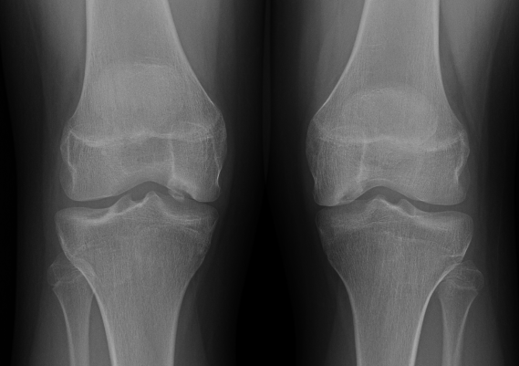
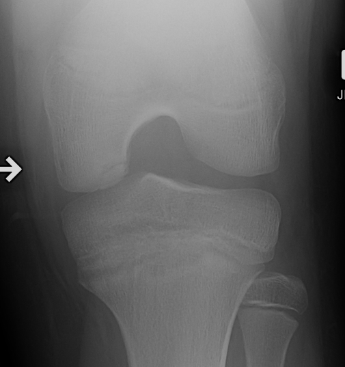
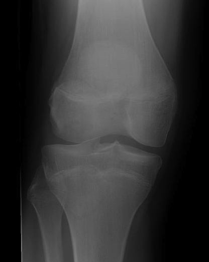
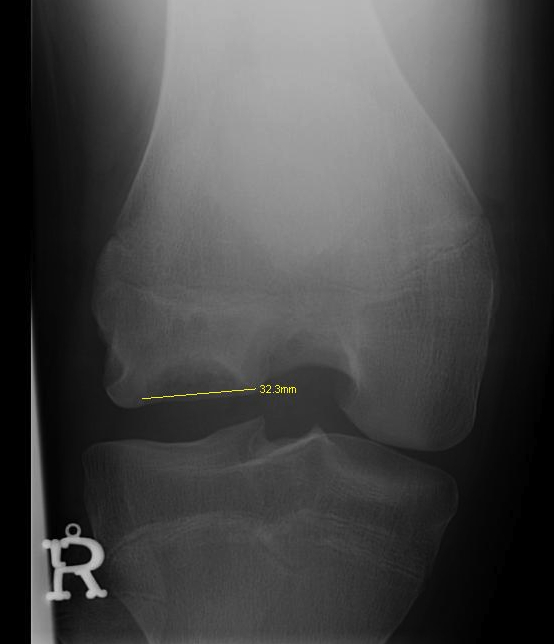
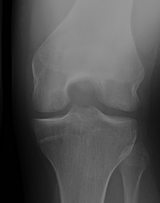
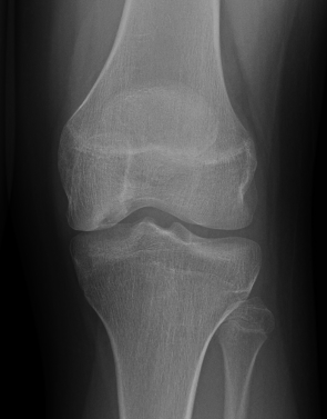
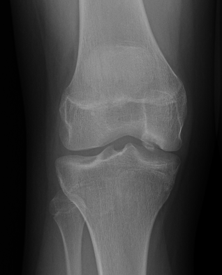
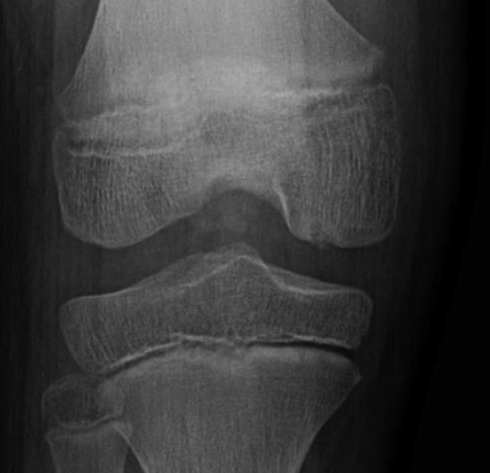
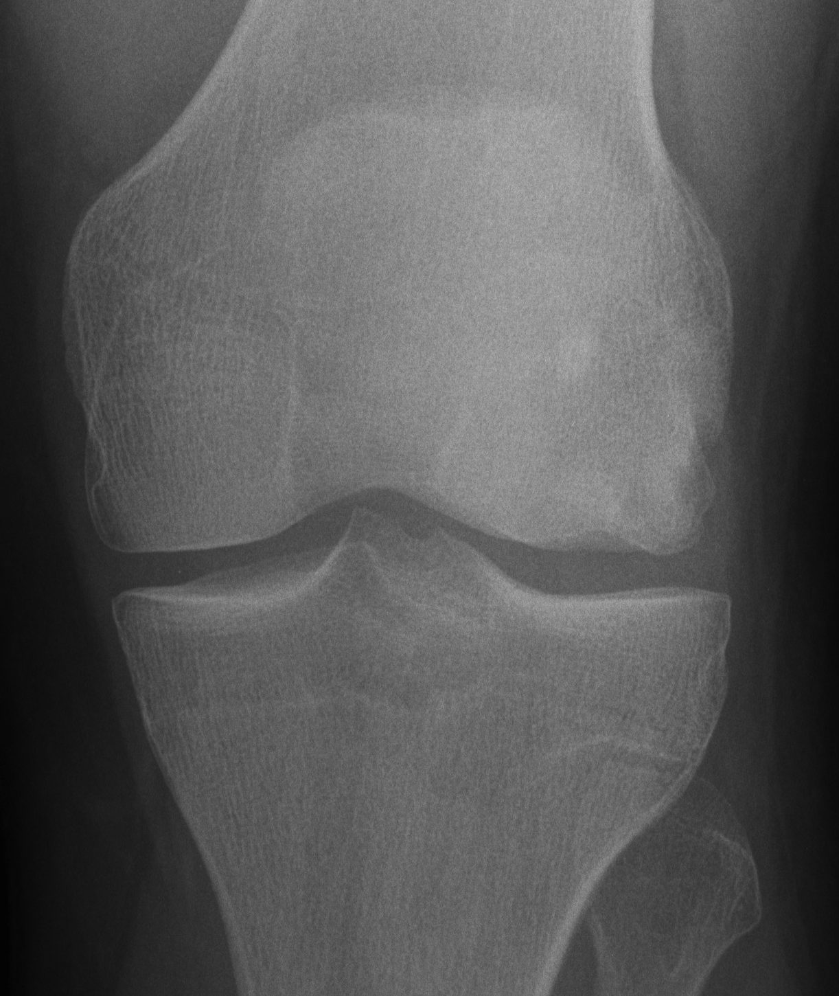
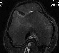
Xray
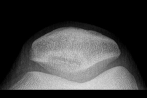
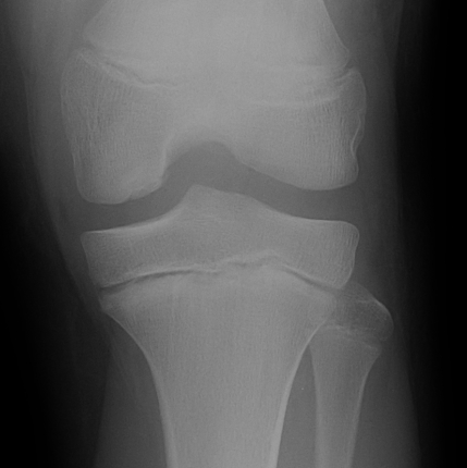
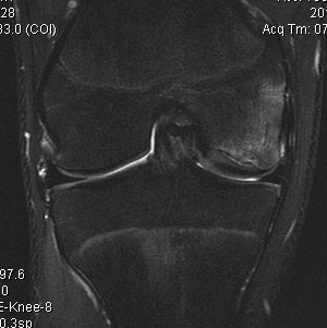
Intercondylar view / notch / tunnel view imperative
- most commonly seen in this view
- can miss the lesion unless have flexed knee view 30-50o
Xray classification
Stage 1: Normal / abnormal MRI
Stage 2: Lucent area of subchondral bone, can have surrounding sclerosis
Stage 3: Partial loosening
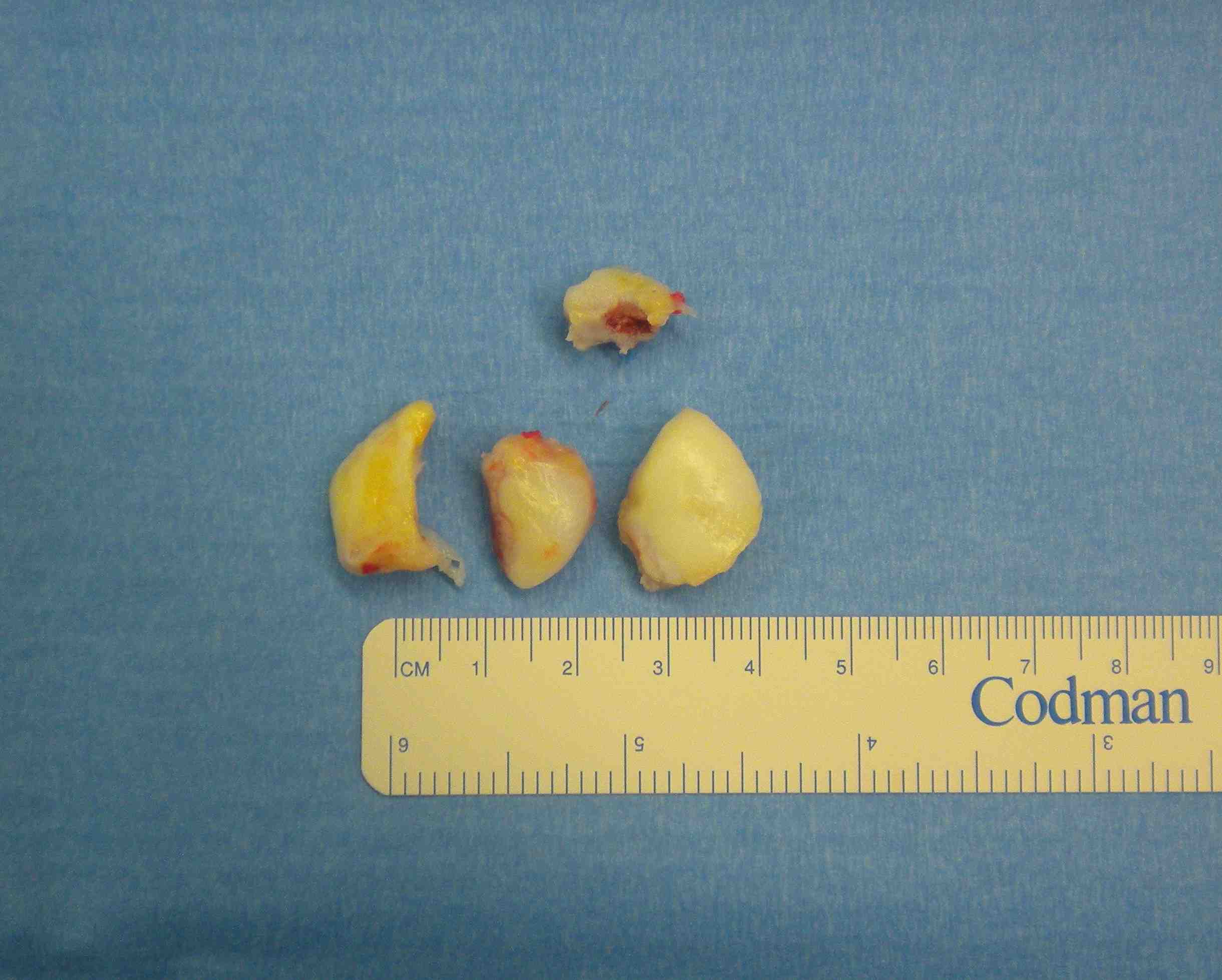
Stage 4: Completely detached / loose body
Type 2 Type 3 Type 3
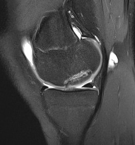
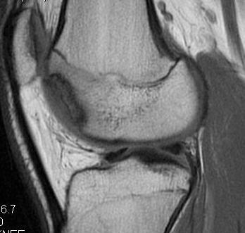
MRI Classification
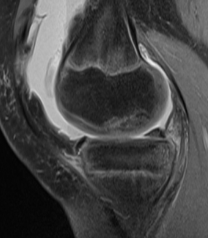
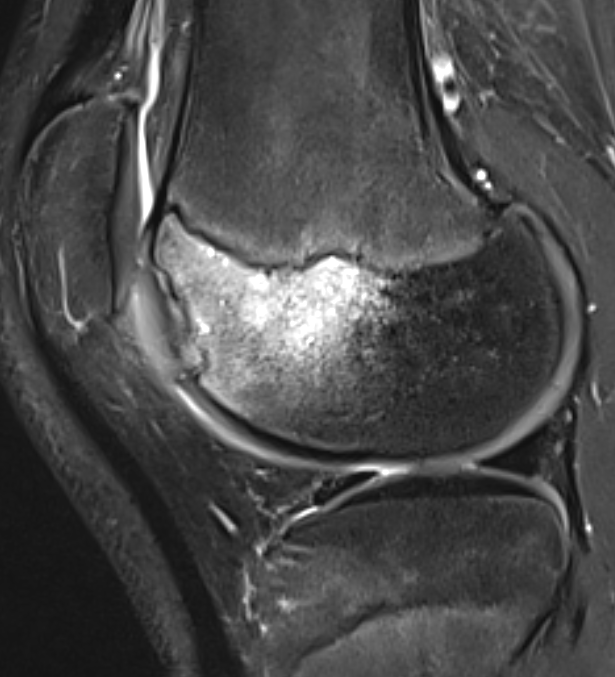
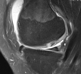
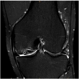
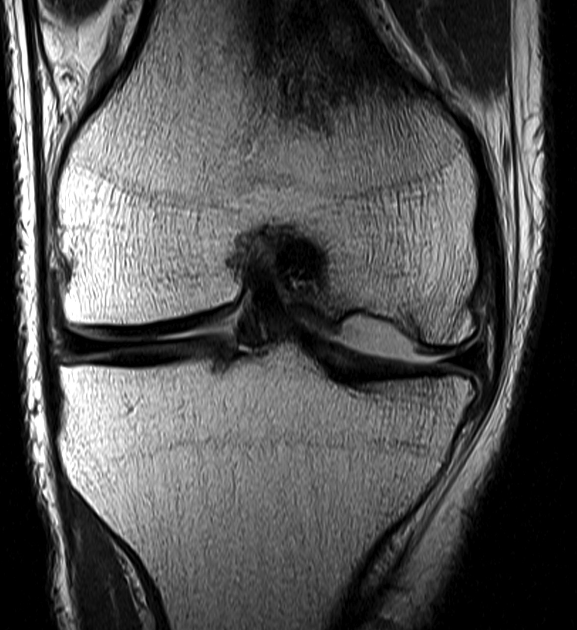
Stage 1: Low signal changes, articular cartilage intact (stable)
Stage 2: Articular cartilage breached, low signal indicating fibrocartilage behind fragment (stable)
Stage 3: Articular cartilage breached, high signal indicating synovial fluid behind fragment (unstable)
Stage 4: Loose body (unstable)
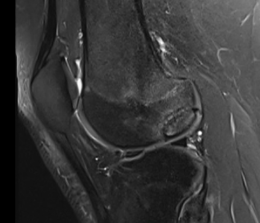
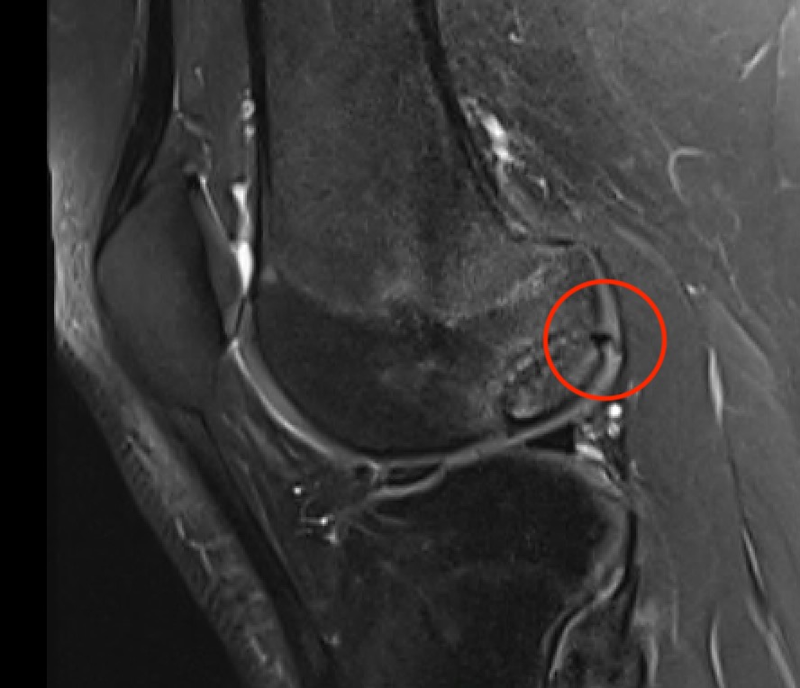
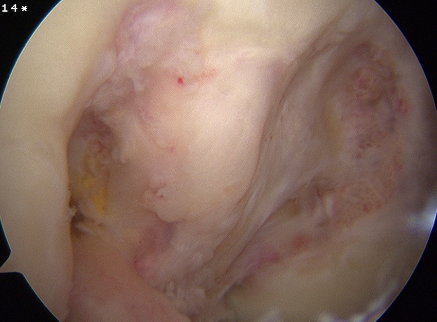
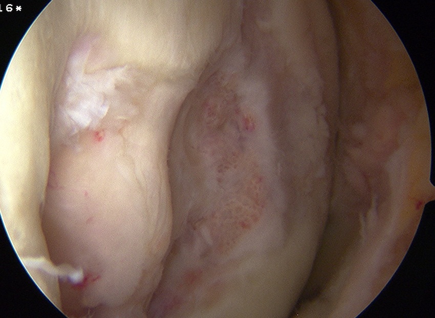
Look for
- integrity of the articular cartilage
- fluid behind the lesion, suggesting instability
- displacement of the lesion
Stable
- no synovial fluid behind lesion
Unstable
- cartilage breach with synovial fluid behind lesion
Stage 1. Articular cartilage intact
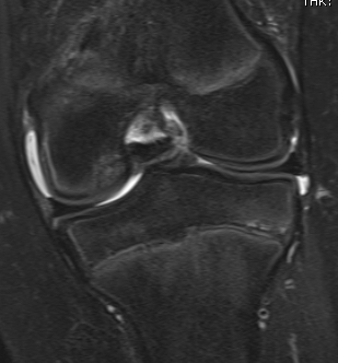
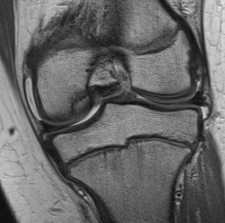
Stage 2. Articular cartilage breach, but low signal intensity behind fragment
Stage 3. Articular cartilage breach and synovial fluid behind fragment (unstable)
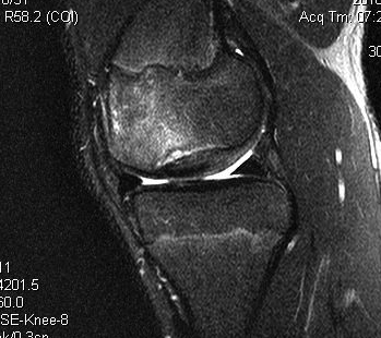
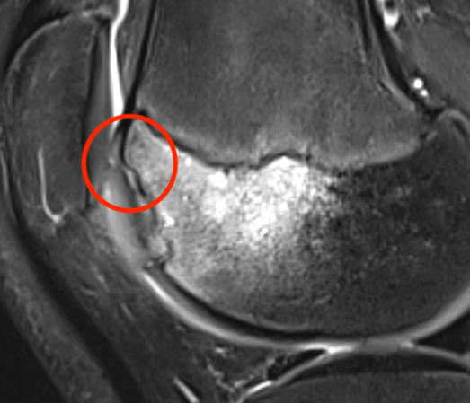
Stage 4. Loose body
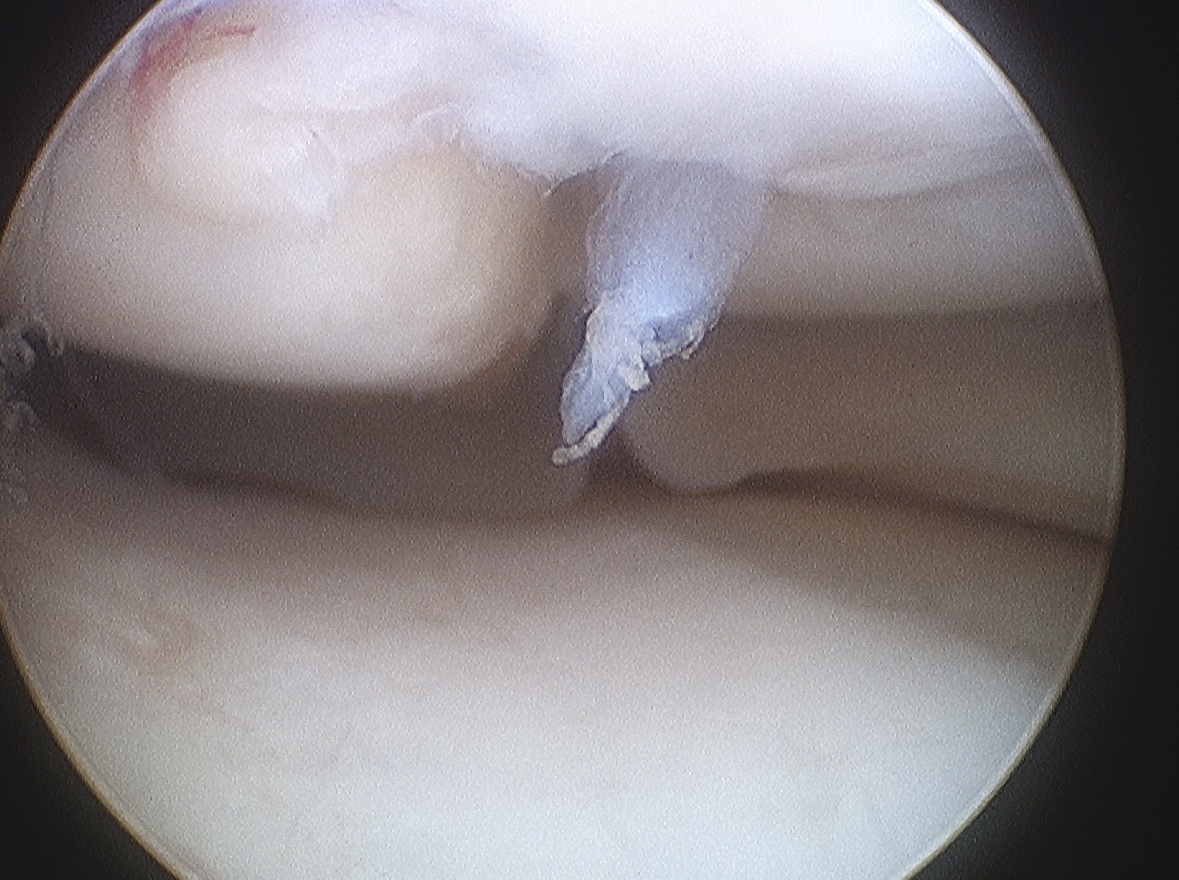
Minimally displaced loose body
Completely detached
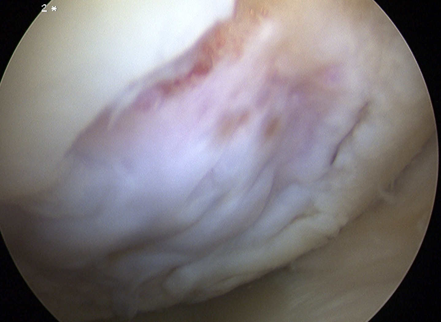
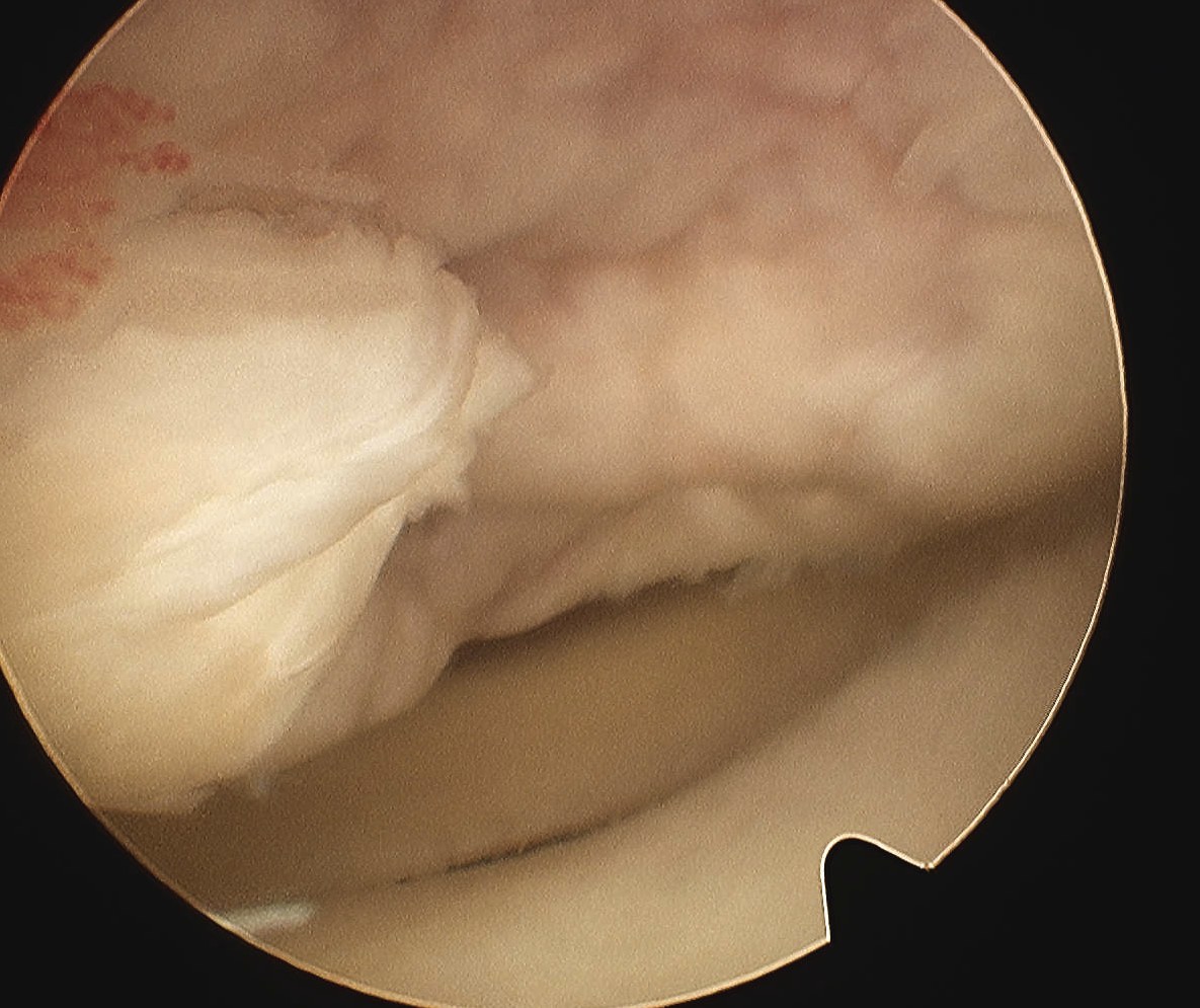
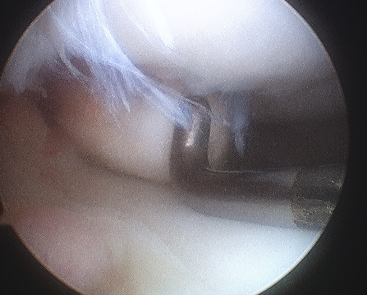
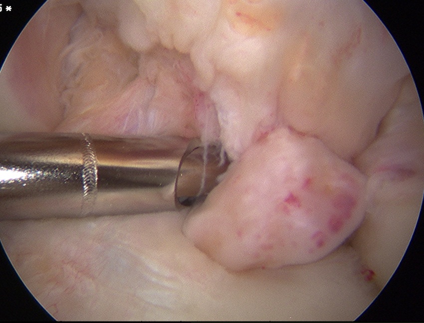
ICRS Arthroscopic Classification
1. Cartilage Intact
2. Partial discontinuity but stable on probing
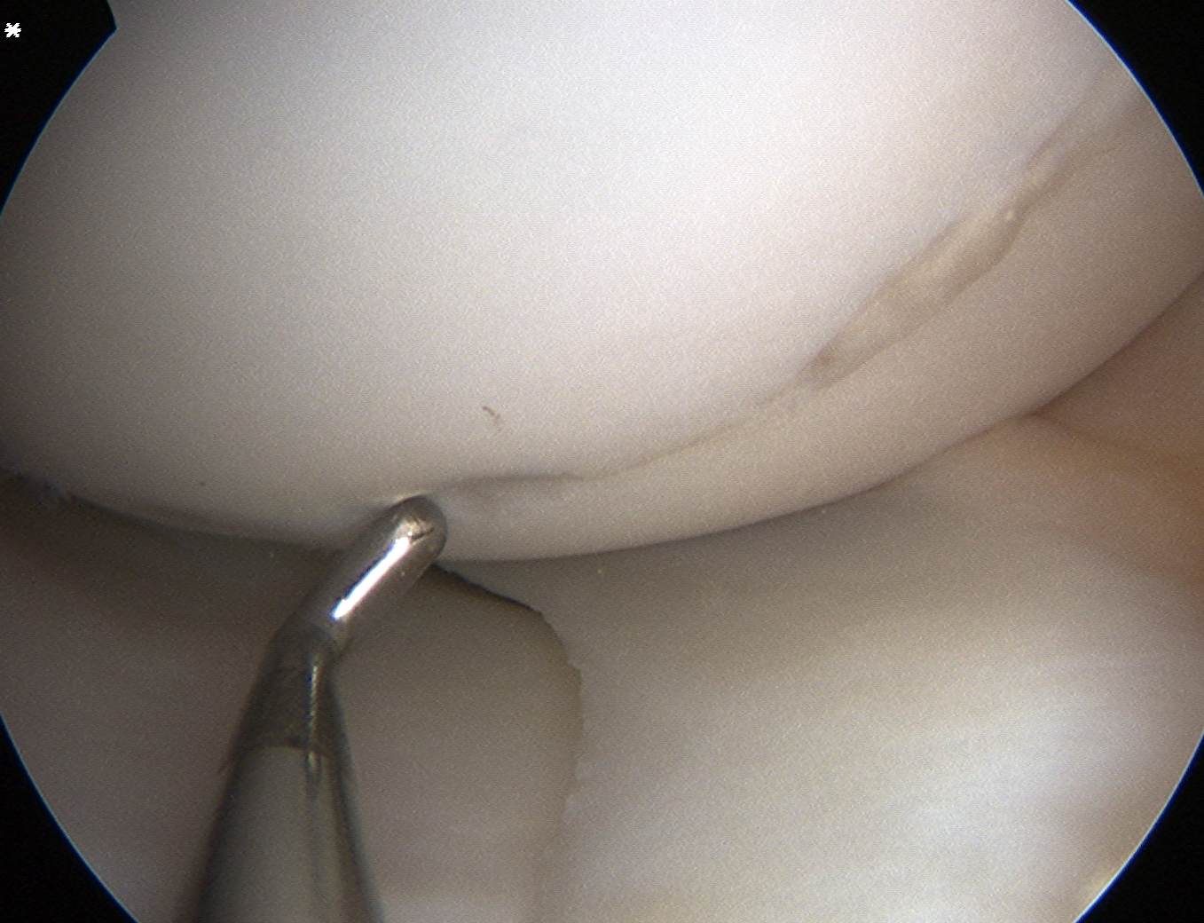
3. Completely detached but insitu
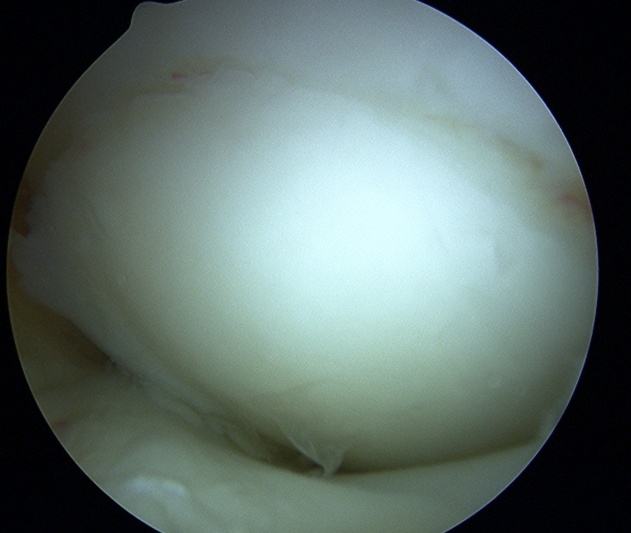
4. Fully detached with crater & loose body
A. Chondral Fragment Salvageable
- recent
B. Chondral fragment unsalvageable
- increased in size / change in shape
MRI and Arthroscopy correlation
Heywood et al. Arthroscopy 2011
- MRI predicted 21/23 OCD to be unstable
- arthroscopy found 10/23 OCD to be unstable
- false positives associated with high signal intensity at bone-fragment interface
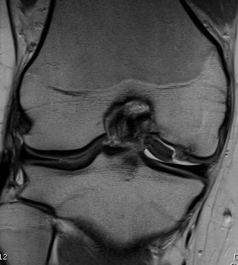
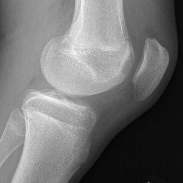
Location
Medial Femoral Condyle 85%
- lateral aspect of the MFC
- PCL origin
Lateral Femoral Condyle 10%
- most common in the central region of the LFC
Patellofemoral Joint 5%
- typically lateral trochlea
Patella OCD