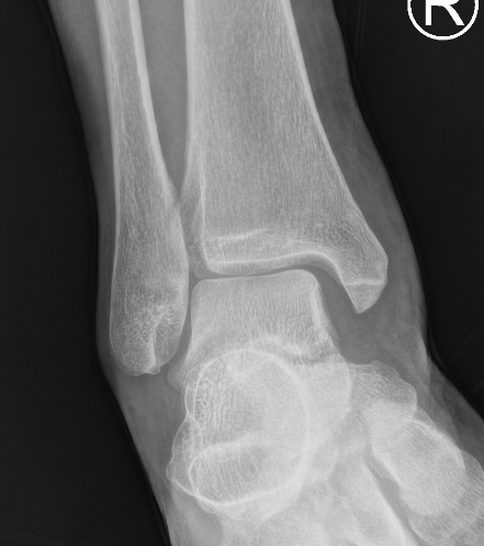
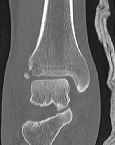
Deltoid ligament injury


Etiology
Ankle sprain
- eversion / external rotation
Ankle fractures


Ankle sprain
- eversion / external rotation
Ankle fractures
1. Posterior Arch
Mechanism
- axial compression with hyperextension
Associations
- 50% incidence other C1/2 fracture
- i.e. ondontoid fracture
Management
- stable
- soft / philadelphia collar
2. Isolated lateral mass fracture
Mechanism
- asymmetrical axial compression / lateral bend
I Open
II Closed
A Supraclavicular
- Preganglionic / Avulsion of Roots
- Postganglionic / Rupture of Trunks
B Infraclavicular
- cords & branches
C. Post anaesthetic
III Radiation / Other
Tumour
Present with pain but not instability
Traumatic
Degenerative
Different treatment algorithms for each
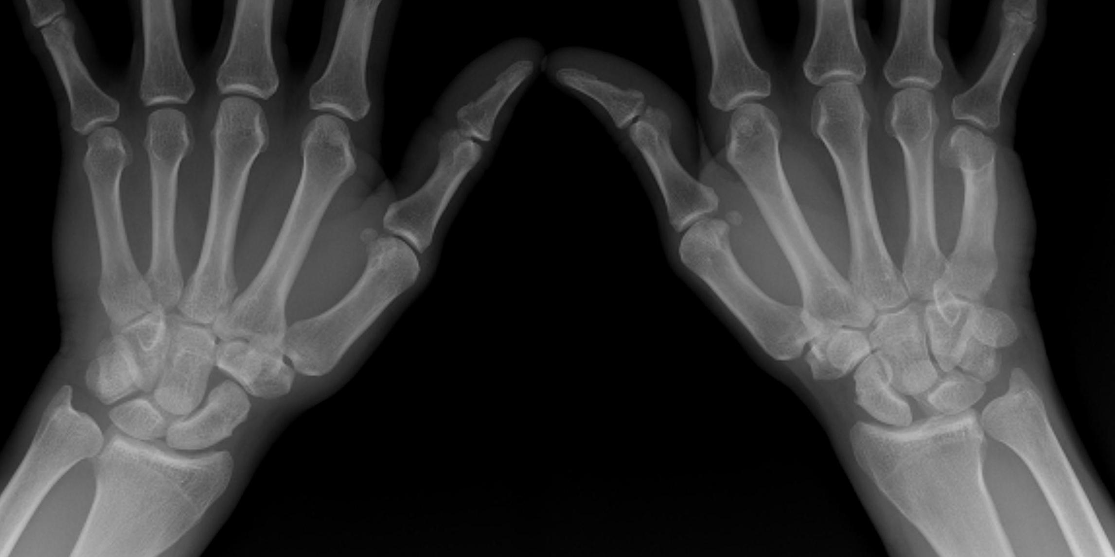
Ulna side wrist pain
- may be worse with rotation
- opening doors and jars
History of trauma
Local tenderness DRUJ

Dorsal Intercalated Segmental Instability / CID
Scapholunate joint
- C shaped
- 2-3 mm thick dorsally with transverse fibres
- thin palmar
Dorsal extrinsic ligaments
- V shaped, onto trapezium
Terminal branch of the posterior cord
- lateral to radial nerve
- behind axillary artery
- runs over inferolateral border of SSC
- enters quadrangular space
Quadrangular space
- SSC superiorly anterior
- T major inferior
- T minor superiorly posterior
- long head triceps and humerus
Divides into anterior and posterior branches

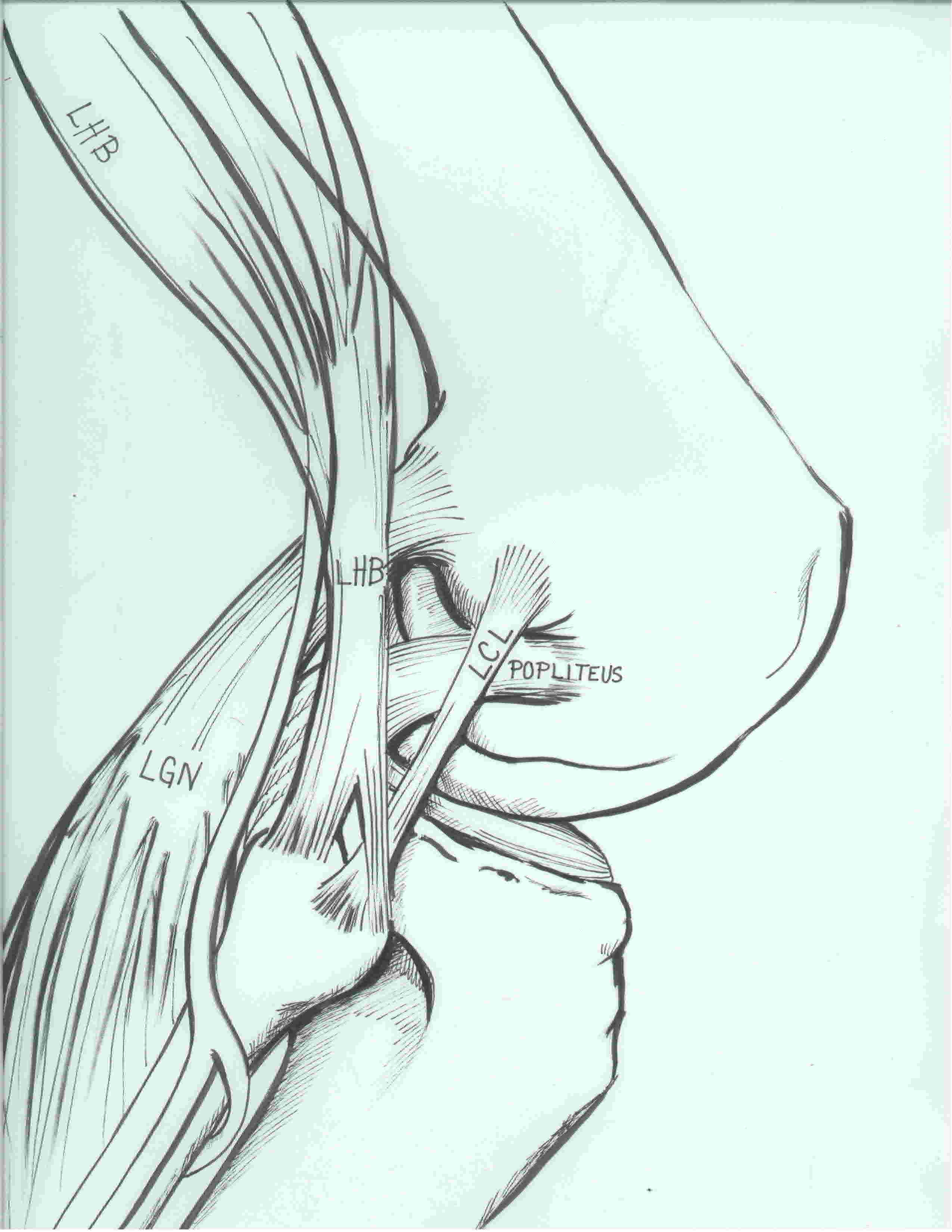
LCL / Popliteus / Popliteofibular ligament
Laprade et al. AJSM 2003
https://pubmed.ncbi.nlm.nih.gov/14623649/
1. Lateral collateral ligament
Femoral attachment
1. Seebacher's 3 layers of the medial knee
Layer 1
- sartorius and sartorius fascia
Layer 2
- superficial MCL
- posterior oblique ligament
- semimembranosus
Layer 3
- deep MCL (meniscofemoral and meniscotibial ligament)
- posteromedial capsule
2. MCL
Superficial MCL
Primary THR 1%
Revision THR 3%
DDH 5%
Sciatic nerve 90% of nerve palsy
Other
- femoral nerve
- CPN
- ulna / radial nerve from positioning
Direct
Laceration
- exposure / sciatic and superior gluteal nerve
- drill reamer / obturator nerve
- spike of cement / obturator nerve