Definition
Benign adipose tumour
Epidemiology
Most common mesenchymal neoplasm
- arise from normal fat
40 - 60 years of age
Clinical
80% subcutaneous
Well circumscribed mobile, round mass
Upper back, shoulder and thigh most common sites
Multiple in 5% of cases
Condition
Durcen's Disease
- multiple lipoma subcutaneous disease
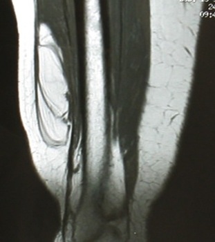
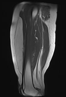
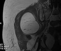
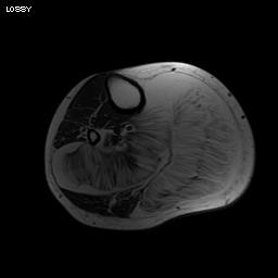
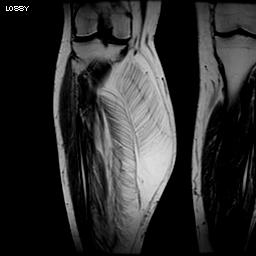
MRI
Same signal intensity as surrounding fat
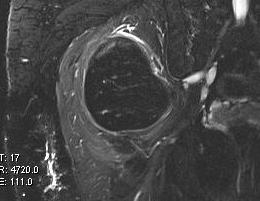
Intra-muscular lipoma
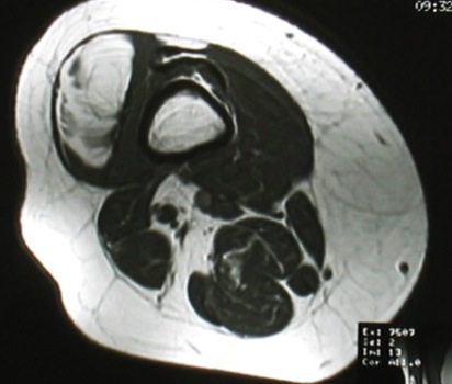
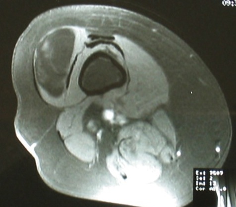
Intra-muscular lipoma
Classification
Divided into 5 subtypes
1. Simple lipoma
2. Spindle Cell Lipoma - variable number of benign spindle cells
3. Pleomorphic Lipoma
4. Intramuscular
5. Angiolipoma - tender / painful
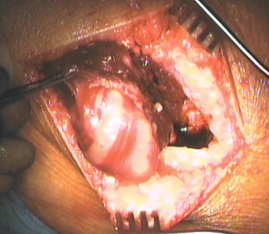
Pathology
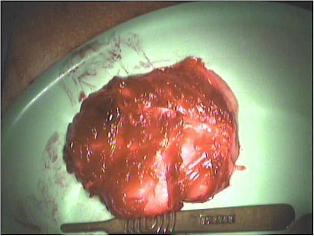
Well circumscribed round to ovoid masses
- homogenous pale to bright yellow on cut surface
Histology
Same as normal fat
- sheets of mature fat cells ovoid to round in shape
- contain single fat droplet with peripheral nucleus
- capillary like vessels are occasionally seen between lobules
Management
Biopsy
Differential diagnosis liposarcoma
Large lesions
Growth
Heterogenous appearance
Deep to fascia
Boneschool page on liposarcoma
Marginal resection
Recurrence uncommon