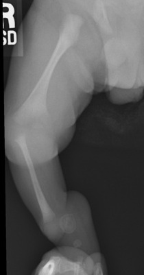
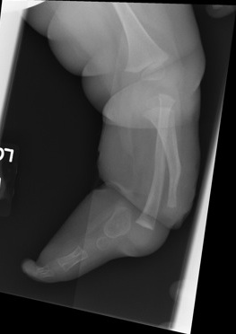
Definition
Paraxial deficiency of skeletal elements on medial aspect of lower limb
Epidemiology
Only skeletal deficiency with a documented familial occurrence
- AD
1/ 1 000 000
Bilateral 30%
Clinical
Leg short +++
Tibial Anterolateral Bowing
Foot fixed in severe varus
- can mimic CTEV
- sole facing perineum
Knee
- FFD
- may be unstable
- no quads mechanism
Associations
Cleft hand
Reduplication of toes
CDH 20%
Classification
MRI to assess extent of proximal failure
1. Unilateral Complete Type 1
No proximal tibial remnant
- usually foot abnormalities
- distal femur is hypoplastic & ossification delayed
- knee is featureless / unstable

Management
- amputation early
- around 1 year before child gets attached to it
2. Unilateral Partial Type II
Well developed proximal tibia & knee joint

Management
A. Knee
- proximal tibiofibular synostosis to prevent proximal migration
- fuse distal fibula to end of tibia
- then either symes or fuse fibula into calcaneus
B. Ankle foot
- distal tibial deficiencies
- get equinovarus deformity similar to club foot
- tibiofibular synostosis
- then either keep foot if good or Symes
3. Bilateral
Management
A. Bilateral through knee amputation
B. Can try to make fibula into tibia and perform symes on one side
- Brown procedure
- need good quadriceps
