Definition
Congenital fibrous, cartilaginous or bony connection of 2 or more tarsal bones
- due to failure of segmentation
Peroneal Spastic Flat Foot
- tarsal coalition
- tarsal pain
- reduced STJ motion
- rigid pes planus
- peroneal muscle spasm / tightness
Epidemiology
Present in 6% of population
- symptomatic in 1% of population
Bilateral in 50%
20% multiple coalitions
AD with variable penetrance
Calcaneo-Navicular most common (2/3)
- talocalcaneal middle facet is next most common (1/3)
- rest uncommon
Associations
Symphalangism (congenital end to end fusion of phalanges)
PFFD
Fibula hemimelia
Other gross limb anomalies
Aetiology
Secondary to failure of differentiation & segmentation of mesenchyme
- supported by intertarsal bridges in fetal tissue
Pathology
Multiple coalitions may occur
- do 2 plane CT with fine cuts to look for other coalitions
- will get poor results if not addressed
May develop ball and socket ankle joint
- due to stiffness of STJ
- develop inversion / eversion in AKJ
Valgus deformity leads to adaptive shortening of peroneal tendons
- may cause reflex spasm of tendon
Classification
1. Location
- CN / TC / TN / CC
2. Ossification
- synostosis - completely ossified
- synchondrosis - partly cartilaginous
- syndesmosis - fibrous
NHx
Majority are asymptomatic & remain so in adulthood
If symptomatic, symptoms usually develop in adolescence when bar ossifies
- due to reduction in STJ movement & joint stress
Calcaneonavicular coalition
- 8 - 12 years of age
Talocalcaneal coalition
- 12 - 16 years of age
History
Present with
- recurrent ankle sprains
- pain over sinus tarsi or over sustentaculum tarsi
- vague aching pain aggravated by activity
Signs
Stiff STJ
- especially talocalcaneal bar
- may still have movement if not ossified
Fixed Pes Planus
- doesn't correct on heel raise or Jack's Test
- heel doesn't swing into varus
- valgus heel with talocalcaneal bar
Peroneal tendons may be shortened but rarely spastic
X-ray
Calcaneonavicular
Oblique xray
- often diagnoses CN bar
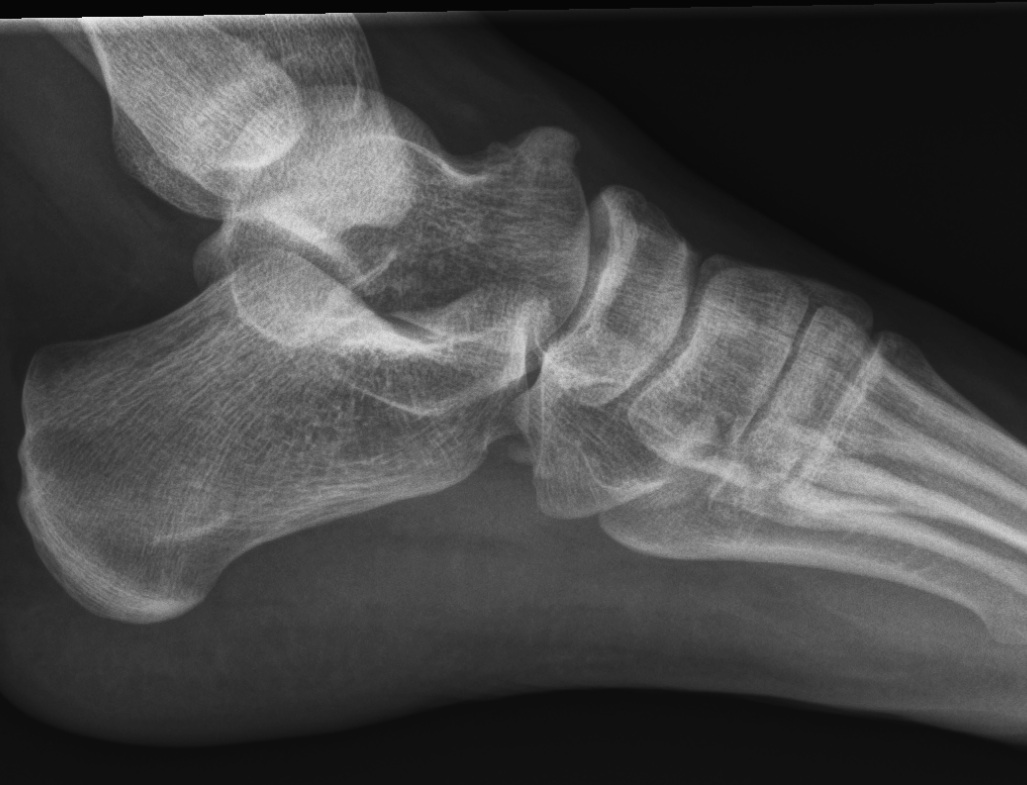
- Anteater sign - elongated process on calcaneus or prolongation of navicular
Talocalcaneal
1. Talar Beaking
- very suggestive of TC bar
- traction spur due to increased stress

2. C sign
- seen on lateral xray
- continuous C shaped line
- from talus to sustenaculum tali
3. Ball and socket ankle joint
- secondary to TC bar
4. Harris axial view
- visualise talocalcaneal
- 40° axial view shows middle facet
- ski jump view
MRI
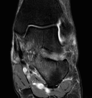
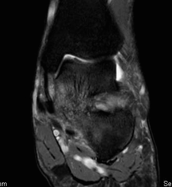
May be helpful for cartilaginous or fibrous bar


CT scan
Very good for bony bars
DDx
Any condition that injures STJ
- traumatic / osteochondral fracture
- inflammatory / RA
- tumour / osteoid osteoma
- infection
Flexible flat foot
Management
Non-operative
Options
Avoid aggravating activities
Moulded longitudinal arch support
SL FWB cast for 6/52
NSAIDS
Operative
Indications
Persistent pain
Minimal degenerative changes
Options
Resection of bar
Isolated STJ fusion - degeneration of STJ only
Triple Arthrodesis - rigid planovalgus foot
Criteria for Resection
All relative
- young < 14 years
- absence of complete bony bar
- no degenerative changes
- presence of talar beaking is not a contraindication
- no fixed deformity
Calcaneonavicular bar resection
NHx compared with talo-calcaneal bar
- possibly settle down long term
- better prognosis / less arthritis / present younger
- more likely amenable to resection
- most do well
Technique
Aim is 1 cm gap
Ollier approach
- 1cm distal to fibular tip
- obliquely across sinus tarsi
- to superolateral margin TNJ
Superficial Dissection
- protect superficial CPN
- EDL & P tertius anteriorly / peroneals plantarward
Deep dissection
- elevate EDB proximal to distal
- beware of its motor branch from DPN
- show sinus tarsi / anterior process calcaneum
- expose bar
- may need to open TN and CC joints to know exact location
Resection
- resect 1cm of bone with osteotomes
- protect talus and STJ from damage
- check with on table oblique lateral II
- suture fat / EDB into defect / over button and felt pad
Post op
- 2 weeks POP
- moon boot / WBAT /ROM
- button out at 6/52
Results
Gonzalez et al JBJS Am 1990
- 75 feet in 48 patients
- good or excellent results in 77%
- poor in 7%
- best results with cartilaginous coalition and patients < 16
Mubarak et al J Pediatr Orthop 2009
- CN resection and fat graft interposition
- 5% incidence of symptomatic regrowth requiring repeat resection
- 74% had improvement of subtalar joint motion
- 82% improvement of plantarflexion
- felt fat graft better choice than EDB as can completely fill gap
Talocalcaneal Bar
NHx
1/3 develop arthritis
- operative results not as successful as CN
- resection more difficult
- results less predictable
- tend to be more conservative with this than CN bar
Prognosis
Favourable
- < 16 years
- < 50% surface area of posterior facet
- nil arthritic changes
- < 16o valgus
Poor
- > 50% of post facet
- i.e. middle facet bar is > 50% of posterior facet
- heel valgus > 16°
- narrowing of STJ i.e. arthritic changes
- lateral talar process impinging on calcaneum
Options
Resection
- is best option if failed non-operative
- always worth trying prior to arthrodesis
- try to return some STJ motion
Calcaneal Osteotomy
- re-centres heel
- realigns weight bearing
- not everyone uses it
Arthrodesis
- in mature foot > 12 years
Resection Technique
Incision
- curved incision
- navicular tuberosity to medial border T Achilles
- 2cm superior to superior calcaneal tuberosity
Superficial dissection
- through flexor retinacular sheath
- elevate T Post and FDL tendon anteriorly
- neurovascular bundle and FHL retracted plantarward
- identify posterior facet
Resection
- resection of bone until middle facet seen + mobile
- remove more bone from talus than sustentacular side
- 2/3 of resection from talus
- key to operation is anterior process of calcaneus & follow posterior
- fat graft or silicon insertion
Post operative
- early ROM important
Talonavicular
Very rare
- ossifies at 3-5 years
- therefore symptoms early
