Aim
Attempt to reduce outliers in all 3 planes of the knee
- improve alignment
- theoretically improve survival and outcomes
Types
Image based
Pre-op CT
- uncommon
- resource heavy
Image less
Anatomic mapping and kinematic analysis of the limb
- intra-operatively
- builds up a working model of the patient's knee
Indications
Unable to use femoral / tibial IM or EM Jigs
- fracture / metal work / deformity
Bilateral TKR
- reduce post op confusion in patient
- from bilateraly IM rod
Young patient < 65
- attempt to achieve perfect alignment / balancing
- want TKR to survive long term
Equipment
2 infra-red trackers
- tibia and femur
- bi-cortical fixation
- must not move intra-operatively
- angled / positioned to be in view of camera
- must be out of way of surgery
Camera / optical localiser
- connected to computer and monitor
Pointer with infrared
Standard cutting blocks
- modified to allow trackers to be attached
Registration
1. Femoral head
- rotate hip whilst minimising pelvic rotation
- kinematic registration
2. Knee
- distal femoral surfaces, epicondylar axis, Whiteside's line
- medial and lateral tibial plateaus, centre of knee, posterior tibial slope
3. Ankle
- medial and lateral malleolus
- ankle centre
Technique
1. Distal Femoral Cut
- leave femoral tracker in place
- cutting blocks attached
- tibial tracker on cutting block
- place at 90o to MA
- set desired resection (depth of femoral implant +/- more if FFD)
- pin block in place and cut
2. Femoral AP cut
- new programs will suggest size
- determines risk of notching
- set rotation from epicondylar axis
- creat drill holes
- place cutting block and cut distal femur AP / chamfers
3. Proximal tibial cut
- reattach tibial tracker
- femoral tracker on cutting block
- cut at 90o to MA
- set posterior slope
- set rotation in line with centre ankle rotation
4. Newer soft ware will assess flexion extension gap
Problems
1. Dependent on registration
- rubbish in = rubbish out
- keep pointer on bone when registering
- accurate landmarks
- movement of pelvis with femoral head rotation
- inherent soft ware inaccuracies
2. May not be as accurate in rotation and sagittal planes as coronal planes
3. Reasons for inaccuracy
- registration
- inaccurate saw cuts (lack of rigid fixation of blocks, width of saw blade)
- uneven cement mantles
Results
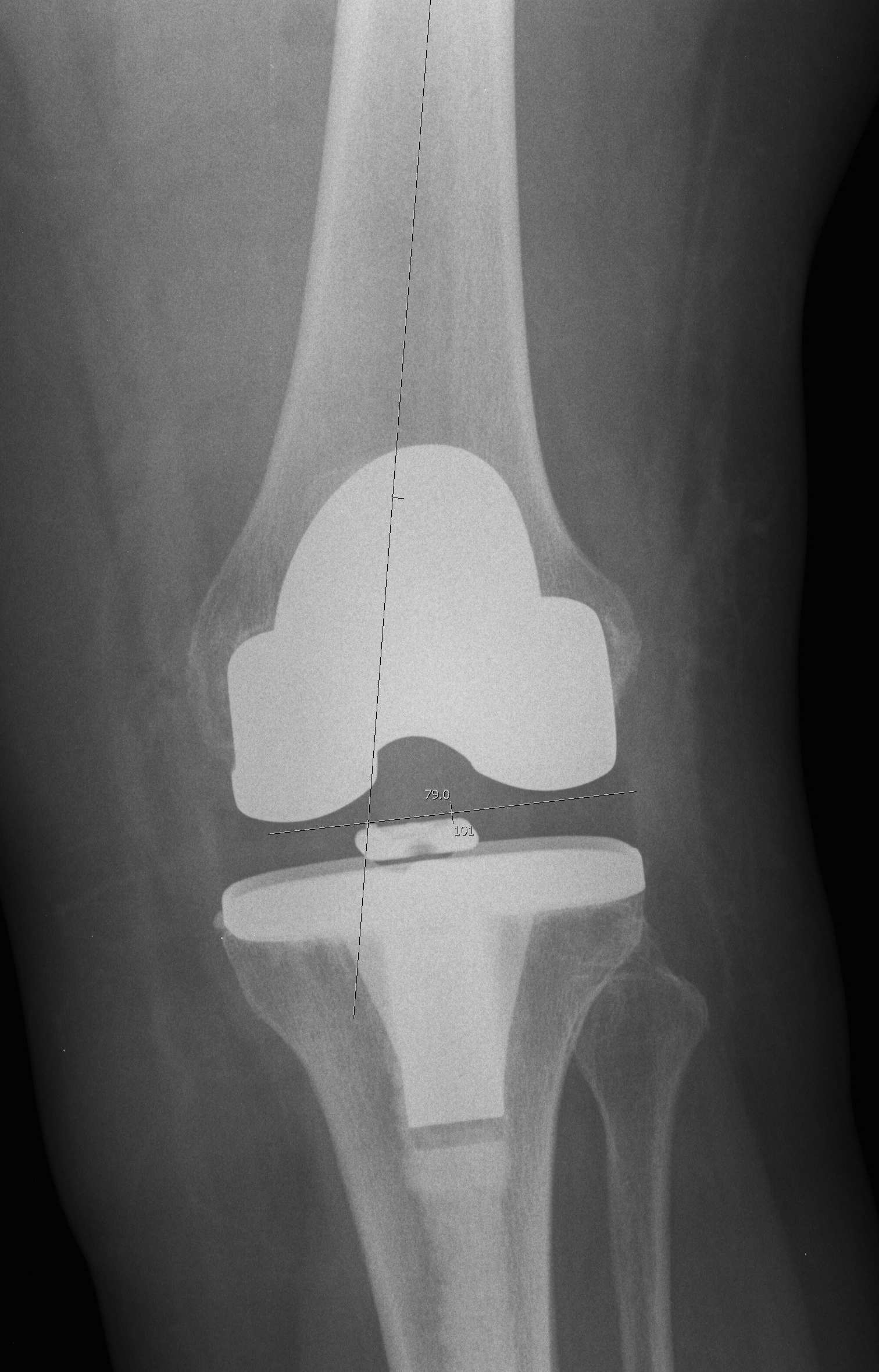
1. Reduces outliers
Will place 90% of TKR's within mechanical axis +/- 3o in coronal plane
- reduces outliers
- will take a long time to see if this improves implant survival
- require large numbers of patients in randomised trials followed over 15-20 years to see a significant result
Lutzner et al JBJS Br 2008
- RCT image less computer navigation
- reduced number of outliers in convential group
- CT analysis showed tibial rotation inaccurate
Spencer et al JBJS Br 2007
- RCT of navigated v conventional
- no difference in functional outcome at 2 years
2. Reduces postoperative confusion
Likely due to lack of IM rod instrumentation
Kalairajah et al JBJS Br 2006
- doppler examination of patients RCT computer v conventional alignment
- significant reduction in emboli in computer navigation
3. Takes longer
Up to 10-15 minutes even with extensive experience
- takes time to insert trackers and perform registration
Bauwens et al JBJS Am 2007
- meta-analysis
- navigation takes 23% longer
4. Reduces blood loss
Kalairajah et al JBJS Br 2005
- RCT
- significant reduction in blood loss in computer navigation group