Definition
Benign Peripheral Nerve Sheath Tumour
- neurofibroma
- schwannoma
Epidemiology
Most common benign nerve sheath tumour
Classification
1. Localised
Most common type
- originate from very small branches of cutaneous nerve
- usually solitary (90%) with no genetic association
- nil malignant transformation
2. Diffuse
Uncommon
- proliferation of spindle cells, collagen & stroma
- permeates subcutaneous tissue & dermis
3. Plexiform
Pathognomonic of Neurofibromatosis Type 1
- massive expansion of nerve by proliferating tumour cells
- small or large nerve
- elephantiasis or gigantism
- 10% malignant transformation
Clinical
Localised
- firm, painless mass
- may have pain / paraesthesia along course of nerve
- + Tinel's
Diffuse - plaque-like cutaneous mass
Plexiform - ropey redundant mass
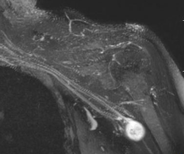
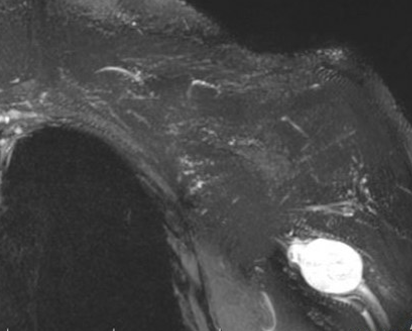
MRI
MRI features of nerve tumours PDF
Target sign
- hypointense centrally
- hyperintense peripherally
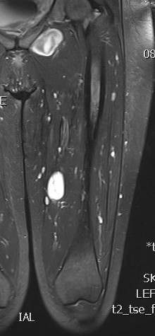
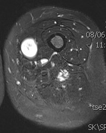
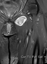
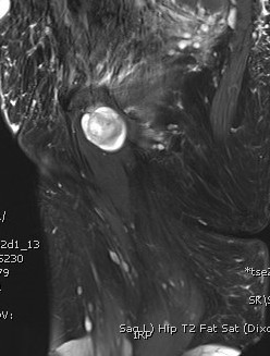
Neurofibroma femoral nerve

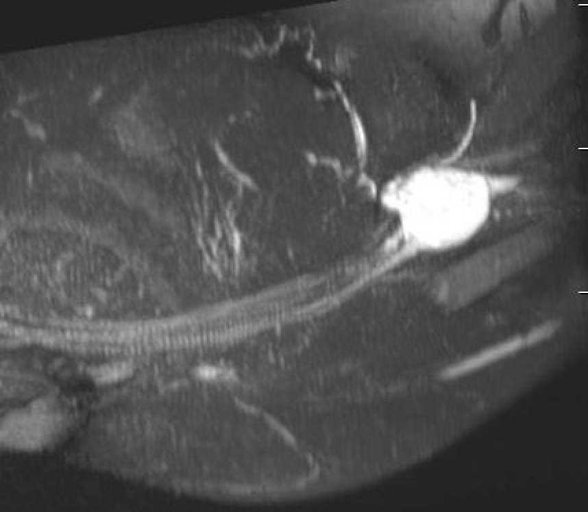
Neurofibroma brachial plexus
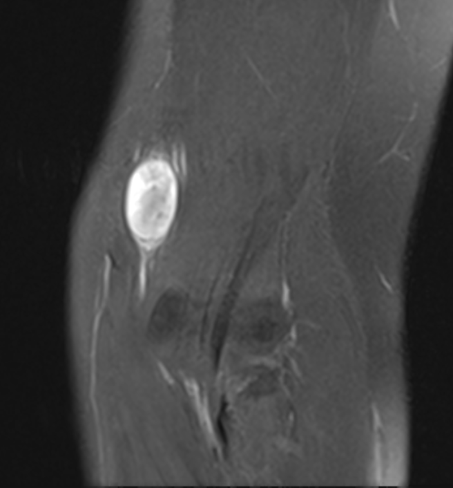
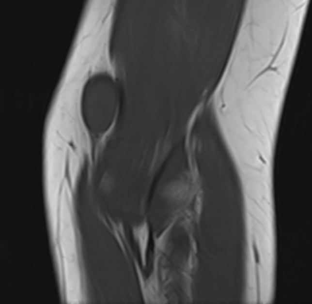
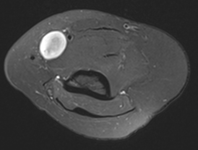
Neurofibroma median nerve
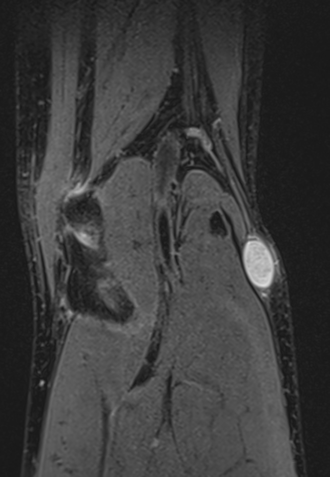
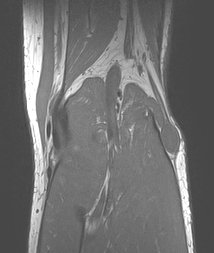
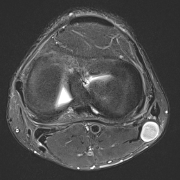
Neurofibroma common peroneal nerve
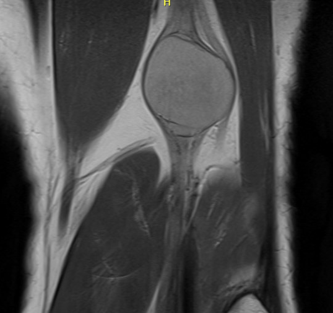
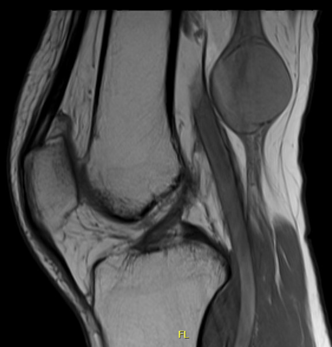
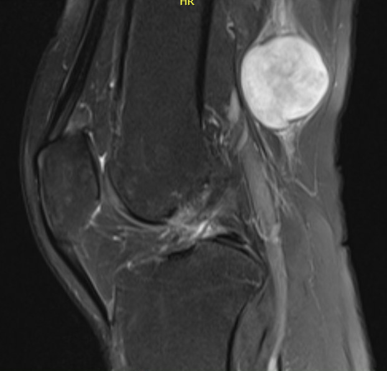
Neurofibroma sciatic nerve
Diagnosis Neurofibromatosis type 1 (NF1) / Von Recklinghausen
(Need 2 of the following)
1. Six or more café-au-lait spots (over 5mm in prepubertal individuals and over 15mm in postpubertal individuals)
2. Two or more neurofibromas of any type or one plexiform neurofibroma
3. Freckling in the axillary or inguinal region
4. Optic glioma
5. Two or more Lisch nodules (iris hamartomas)
6. A distinctive osseous lesion such as sphenoid dysplasia or thinning of the long bone cortex with or without pseudarthrosis
7. A first degree relative (parent, sibling, or offspring) with NF1 by the above criteria
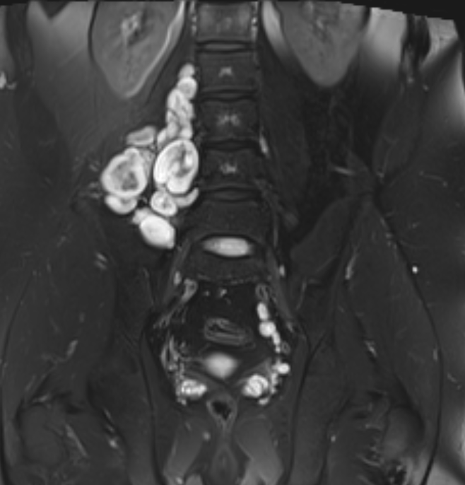
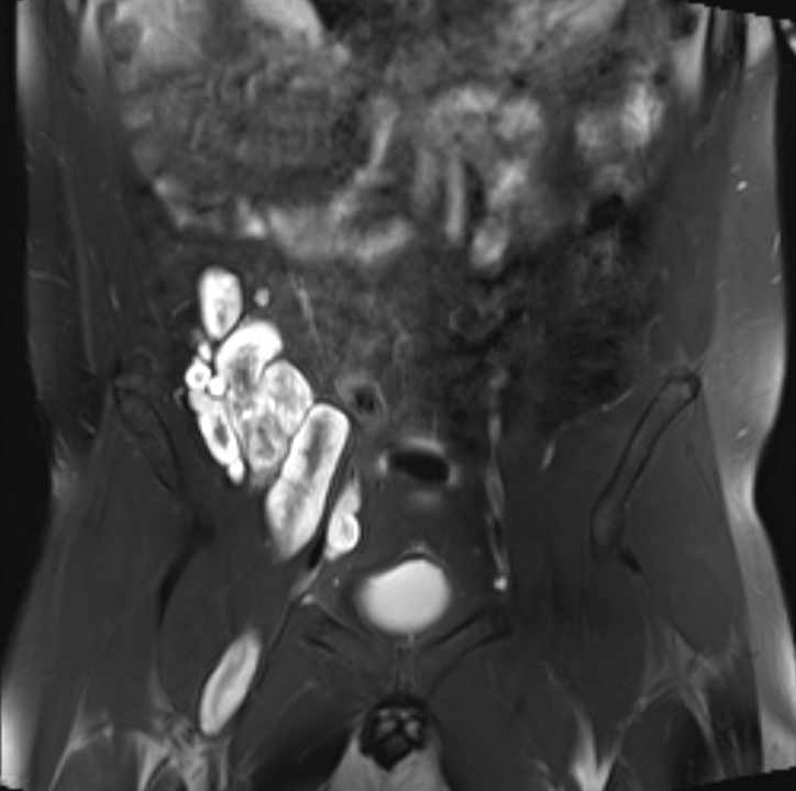
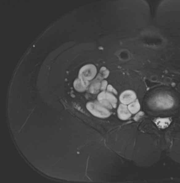
Plexiform neurofibromas Neurofibromatosis Type 1
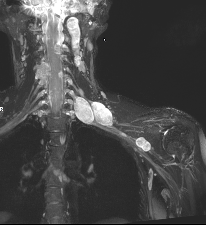
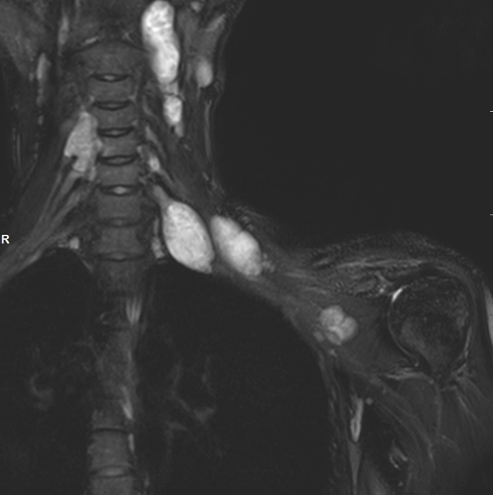
Multiple neurofibromas Neurofibromatosis Type 1
Histology
Neurofibroma - proliferation of schwann cells / perineural cells / fibroblasts
Schwannomas / neurilemmoma - contain only Schwann cells
Management
Benign Peripheral Nerve Sheath Tumour
Technique
- not easily separated from nerve
- excision usually involves partial resection of nerve
- cutaneous nerve: resection
- nerve large and important: need interposition graft
Malignant Peripheral Nerve Sheath Tumour
Indications for biopsy
- rapid growth
- night pain
- size > 5 cm
Epidemiology
- 10% of sarcomas
- 50% seen in neurofibromatosis
- NF1 - 50% lifetime risk
Site
- brachial plexus
- lumbosacral plexus
- major nerve trunks - especially the sciatic nerve
MRI
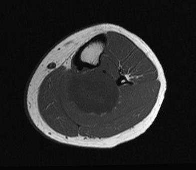
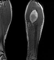
Management
Aggressive, malignant tumours
Wide resection + radiotherapy
Kolberg et al Neuro Oncol 2013
- meta-analysis of 1,800 patients
- overall survival 44%
