Bursas
12 in total
- 4 anterior
- 4 lateral
- 4 medial
Anterior
Suprapatellar
Prepatellar
Superficial & Deep Infrapatellar
Lateral
Biceps femoris - between biceps & LCL
LCL - between LCL & capsule over popliteus tendon
Lateral Gastrocnemius - between LG & capsule
Popliteus - between popliteus tendon & tibia & fibula
Medial
Pes anserine - between pes & MCL
MCL - between MCL & tibia/capsule/SM radiation
Semimembranosus - between SM & MG
Medial Gastrocnemius - between MG & capsule
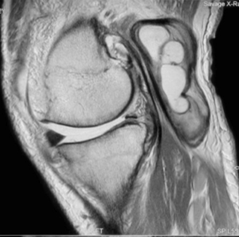
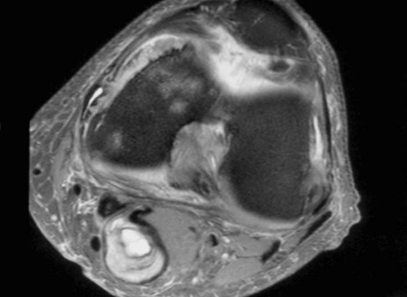
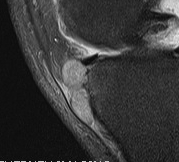
Popliteal Cyst / Baker's Cyst
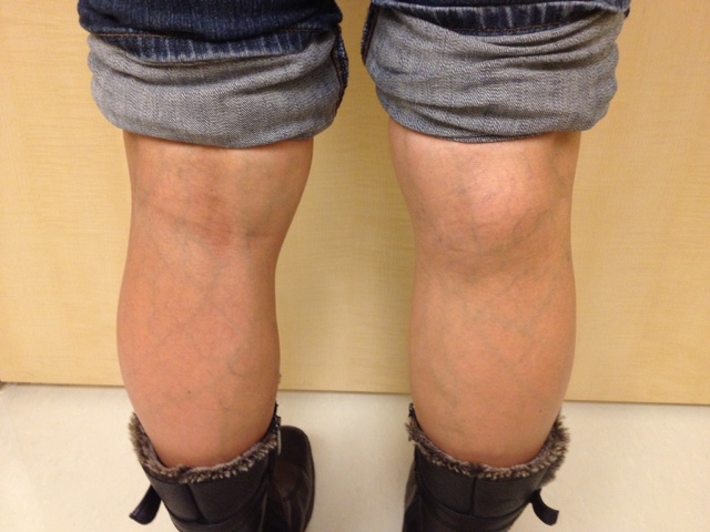
Semimembranosus bursae or medial gastrocneumius
- can be herniation of synovium through capsule
- increases with fluid in the knee / OA / inflammatory conditions
- one way valve
Site
In midline and below joint line
- position crucial to avoid confusion with ST tumour
Pathology
May leak or rupture
- can be very painful
- causes swollen tender calf & mimics DVT
MRI
Diagnosis
- must communicate with knee joint
DDx of Popliteal Mass
1. Bakers cyst
- transilluminate
- mobile and soft
- medial and distal to flexor crease
2. Lipoma
3. Aneurysm
- pulsatile
4. ST Tumour
- rhabdomyosarcoma / synovial hemangioma / PVNS
- hard, fixed, don't transilluminate, calcification
Semimembranosus Bursa
Pathology
Enlargement of bursa
- presents betwen semimembranosus & head MG
- occurs in children & young adults
Presentation
Painless lump behind knee
- inverted U shape
- medial to midline
- most prominent with knee straight
Knee joint is normal
- lump may ache
Management
Usually resolves after 1-2 years
Excision should be avoided
- 2/3 communicates with knee
Popliteal Cyst in children
Very common
- medially, distal to flexor crease
- may be associated with JCA or PVNS
Management
Resolve over 10-20 months
- 50% recurrence with excision
- do not operate
Prepatellar Bursitis / Housemaid's knee
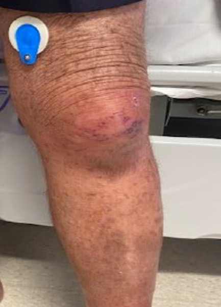
Aetiology
Due to friction between skin & patella
- occurs with repetitive kneeling
Clinical
Circumscribed fluctuant swelling anterior to patella
- knee joint normal
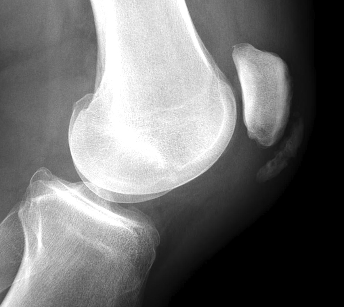
Xray
May see calcification in long standing cases
Management
Rest / knee pad
- may need excision if recurrent & troublesome
Infrapatellar Bursitis / Clergyman's knee
Similar to prepatellar bursitis
- swelling superficial to PT
- more distal
Pes Anserinus Bursitis
Clinical
Occurs over medial upper tibia
- deep to sartorius, gracilis, semiT
- lies between pes anserine & MCL
Pain and tenderness over insertion
Xray
Exclude bony pathology
Biceps Femoris Bursa
Related to biceps insertion into fibula head
- may be confused with ganglion from superior tibio-fibular joint