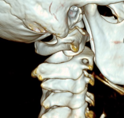
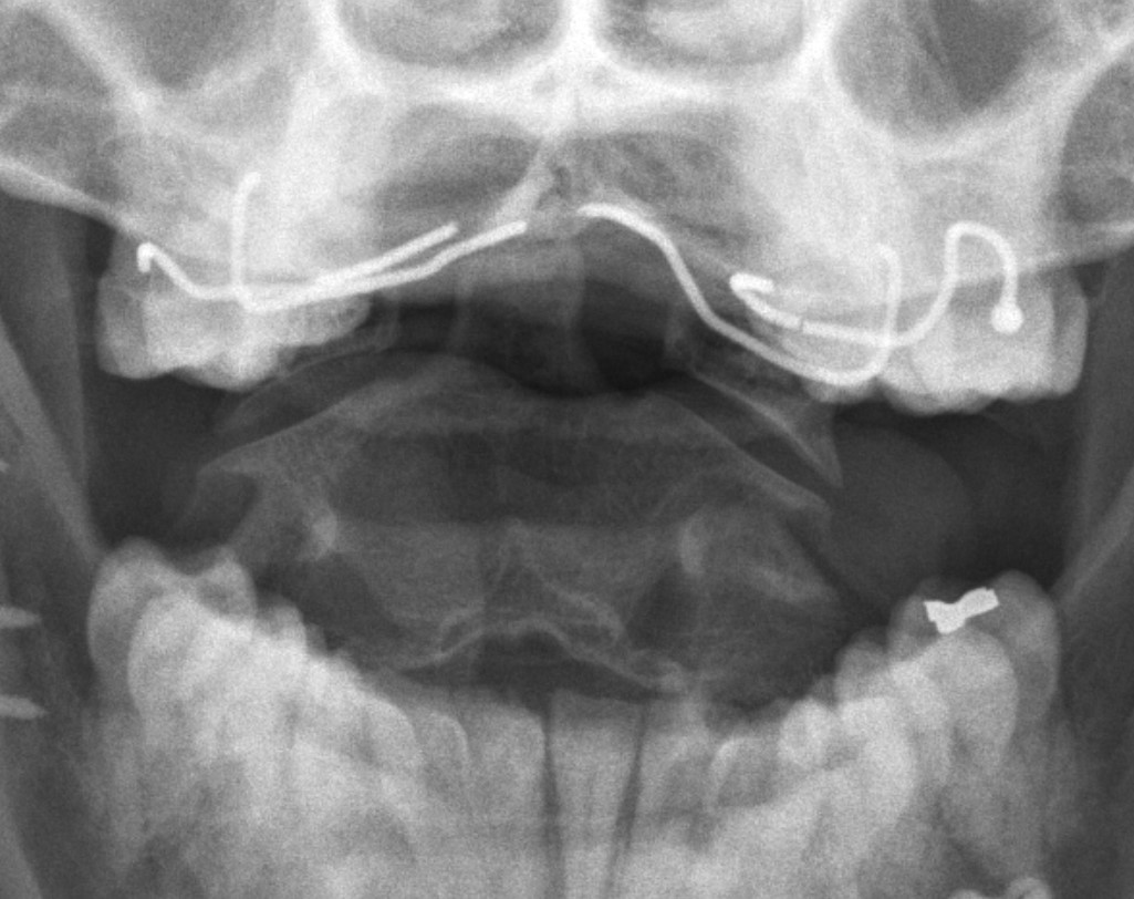
anatomy
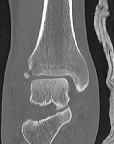
Posterior process fractures
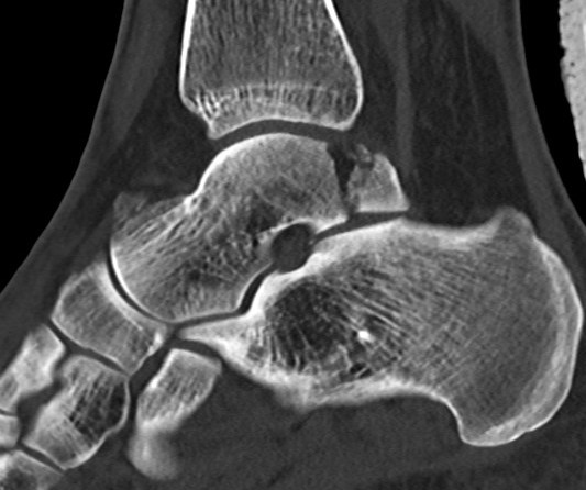
Anatomy
Posterolateral & Posteromedial tubercles
- separated by sulcus for FHL
- lateral larger than medial
PL tubercle
- size variable
Deltoid ligament injury
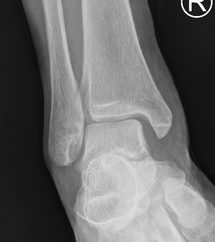
Etiology
Ankle sprain
- eversion / external rotation
Ankle fractures
Cord injury patterns
Anatomy
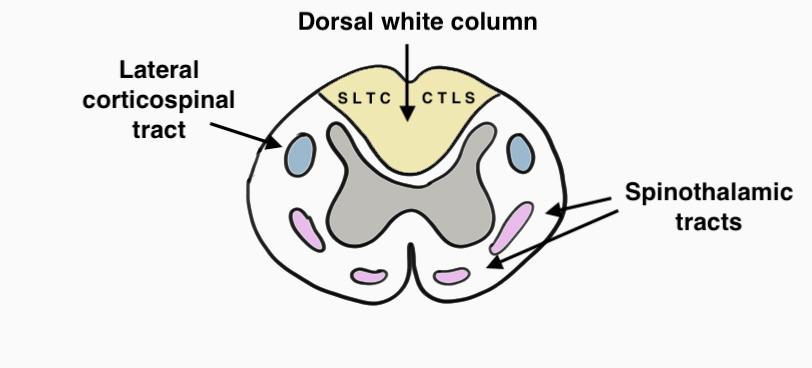
Dorsal Columns
- light touch, vibration & proprioception
- CTLS (cervical fibres central, sacral fibres lateral)
Lateral Corticospinal Tract
- motor tract
Ulna collateral ligament
Definition
Injury to ulnar collateral ligament of thumb MCPJ
Interferes with pinch grip and grasp and thumb is ineffective as a post


Pectoralis Major Tears
Epidemiology
Middle age men
Steroids / Growth Hormone
Aetiology
Usually occurs in gym
Bench Press
Clinical
Significant bruising in the acute phase
In chronic setting, ask patient to adduct against hip / resistance
Arterial Supply Upper Limb
Subclavian Artery
Origin
- on right arises from brachiocephalic trunk behind right sternoclavicular joint
- on left arises from the arch of the aorta
Ends
- outer border first rib as axillary artery
First part
- origin of artery to medial aspect scalenus anterior
- on right arches above the clavicle / lies on the pleura / RLN wraps about it
Arterial Supply Lower Limb
Femoral Artery
Enters thigh
- midway between ASIS and pubic symphysis
- in femoral triangle
- NAVY (nerve / artery / vein / Y fronts)
- in femoral sheath with femoral vein (transversalis fascia and psoas fascia)
- femoral nerve outside sheath / under the iliac fascia / lateral
Femoral triangle
Anatomy
- inguinal ligament superiorly
- medial border sartorius laterally

