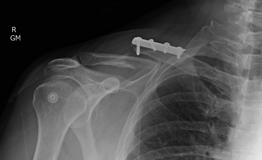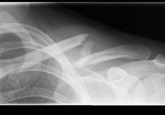
Mechanism
Usually a direct blow
- less commonly a fall on the outstretched hand
RTA / sporting accidents commonest causes
Can be pathological as a result of radionecrosis
- eg following radiotherapy for breast cancer.
Incidence
Fractures of the clavicle are common
- 5% of all fractures
- Up to 80% involve the middle third
Anatomy
Ossification
First bone to ossify in 5th week of foetal life
- intramembranous ossification
- medial growth plate accounts for 80% length
- medial physis last to close at 22-25 years
Shape
The middle third of the clavicle is the junction of two curves
- medial convex anteriorly
- lateral convex posteriorly
At the junction there is little cancellous bone
- skeletal muscle covers only part of the cortical bone
- the volume of muscle in this region is small
The clavicle is secured firmly at each end by stout ligaments and joint capsules
Movement
It rotates approximately 40o when the scapula is elevated
- most of the rotation occurring after the arm passes the horizontal level
Classification
Fractures may be divided into three regions of the clavicle
Medial end
- fifth of the bone
- lying medial to a vertical line drawn upward from the center of the first rib
- rare
- <5%
Lateral clavicle fracture
- fifth of the clavicle
- lateral to a vertical line drawn upward from the center of the base of the coracoid process
- a point marked by the conoid tuberosity
- approximately 1/3 of all clavicle fractures
Diaphysis
- intermediate three-fifths between these two areas
- most common
- 70% of clavicle fractures
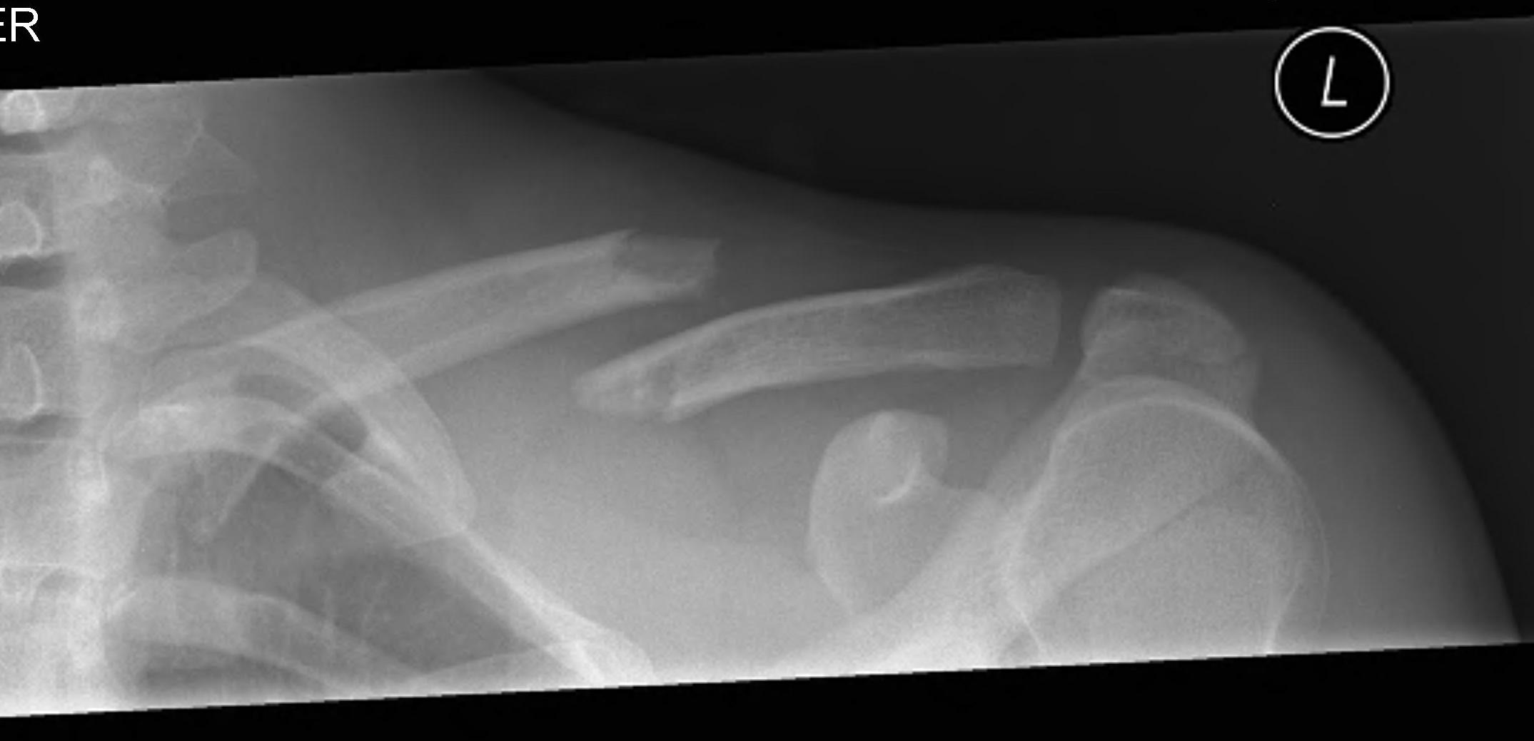
Examination
Examine skin
- ensure skin not threatened by spike of bone
Examine AXN
- sensation in deltoid patch
Look for scapula winging
- may be an indication for fixation
Xray
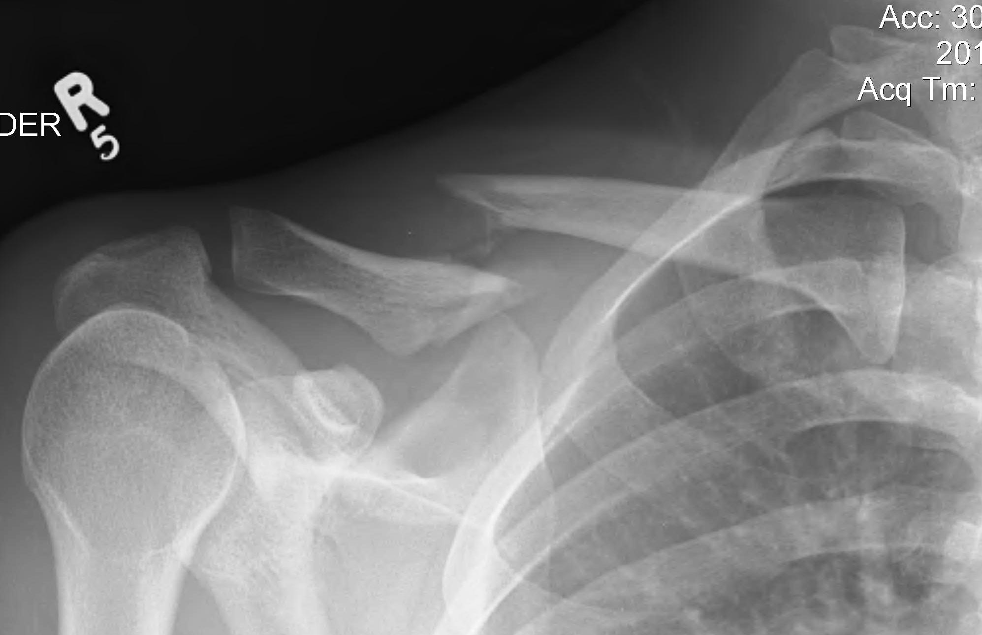
Displacement
- the proximal end under the pull of SCM becomes elevated
- the shoulder tends to sag downwards and forwards
- with further displacement there is overlapping and shortening
- the portions of the clavicle may also be rotated relative to one another
Non Operative Management
Sling for comfort
- followed by early mobilisation as the pain subsides
Figure of 8 bandage
- these do not effectively reduce the fracture
Complications Non Operative Management
1. Persistent bony spike
- even after normal remodelling
- may require excision
2. Thoracic outlet syndrome
- secondary to hypertrophic non-union of the clavicle
- also due to reduced subclavicular space in a shortened malunion
- late compression of ulna nerve, brachial plexus
- symptoms with overhead activities
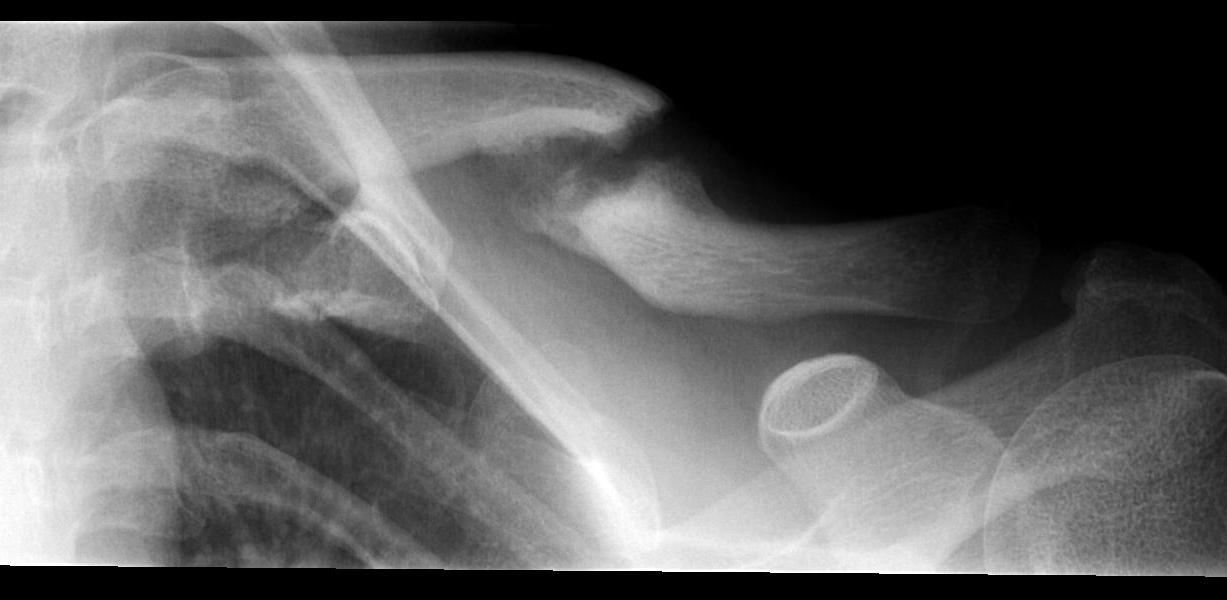
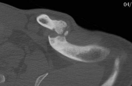
2. Non union
Neer (6) 1960 JAMA
- 2235 closed clavicle fractures treated non-operatively
- non union in only 3 (0.1%)
- his series of 45 fractures treated by open reduction, there was non union in 2 (4%)
Rowe Clin Orthop 1968
- nonunion in 0.8% of fractures treated by closed methods
- 3.7 % in those treated by open reduction
Robinson et al JBJS Am 2004
- prospective cohort study
- overall non union rate in clavicle fractures of 6.2%
- 4.5% of diaphyseal fractures / 11.5% lateral fractures / 8.3% medial fractures
- factors increasing risk non-union in diaphyseal fractures
- advancing age / female gender / complete displacement / comminution
3. Malunion

Hill et al JBJS Br 1997
- studied 52 completely displaced midshaft clavicle fractures
- all treated non operatively
- 8/52 (15%) developed non union
- 16/52 (31%) unsatisfactory (residual pain / brachial plexus symptoms)
- initial shortening of > 20 mm associated with non union and poor outcome
McKee et al JBJS Am 2003
- poorer functional outcome
- fractures > 2cm shortening
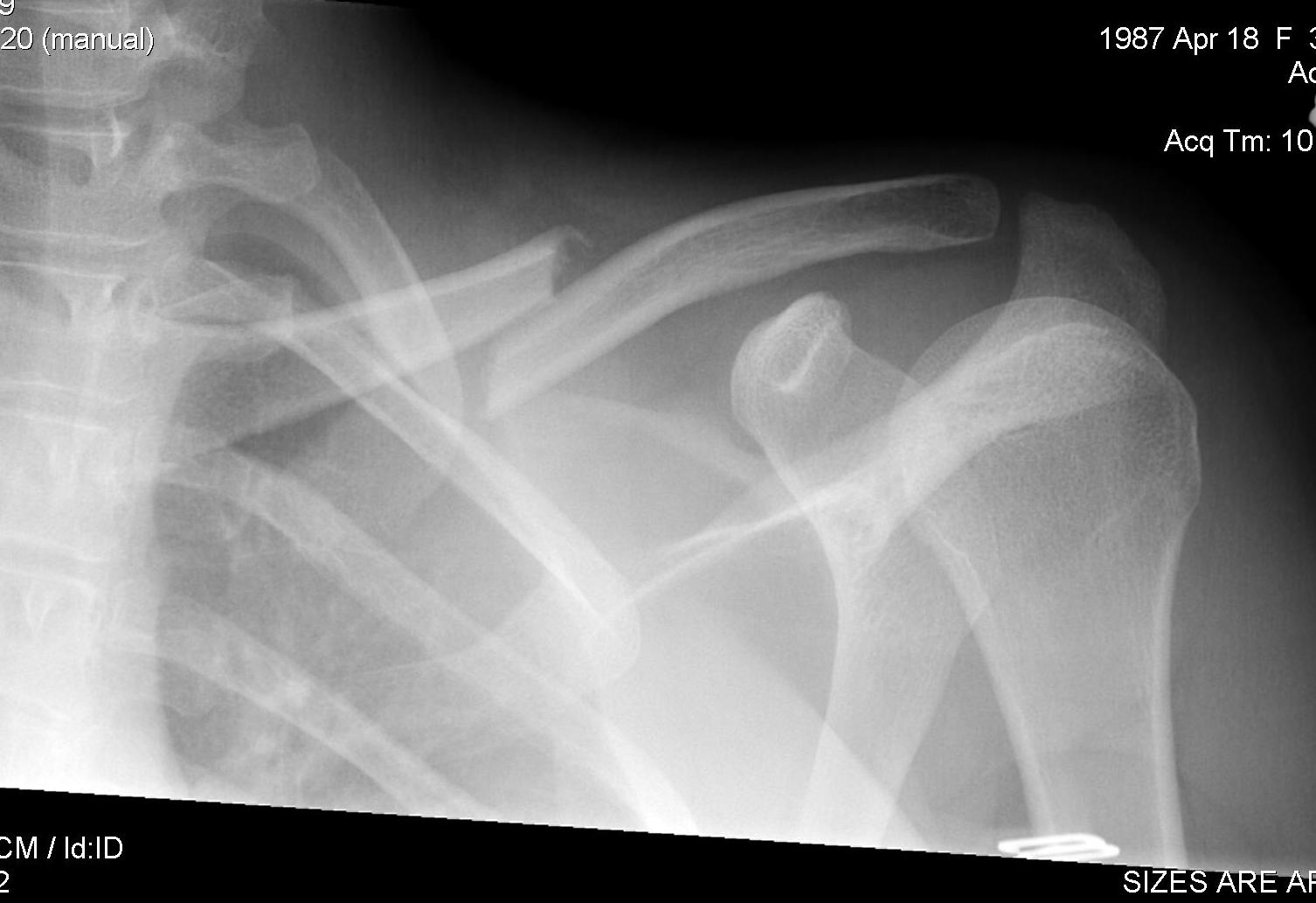
Canadian Orthopedic Trauma Society JBJS Am 2007
- multicentred RCT of op (62) v non op (49) treatment displaced midshaft clavicles
- 1 year followup
- average time to radiographic union 28 v 16 weeks
- non unions 2/62 v 7/49
- symptomatic malunion 0/62 v 9/49
- better shoulder scores at all times
- in operative group, 3 wound infections, 1 mechanical failure and 5 prominent hardware
Operative Management
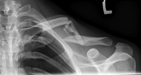
Absolute indications
Skin compromise (open fracture or severe skin tenting)
Neurovascular injury
Relative indications
Floating shoulder
Multi-trauma
High risk of non union / malunion
Non union
1. Plate fixation
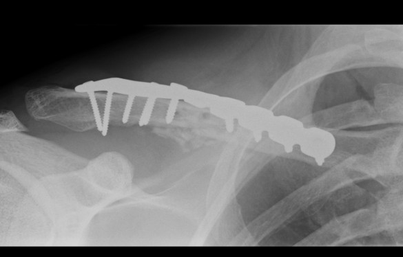

Indications
- fresh fractures of the middle third
- gross displacement and angulation of the bone
- shortening of the clavicle that was estimated to exceed 2.5 cm on plain radiographs
Results
Bostman et al J Trauma 1997
- plate fixation (DCP / Pelvic Recon)
- ORIF in 103 (9.5%) of the total of 1081 patients
- complication rate 23%
- infection rate was 7.8%
- both patients treated with a 1/3 tubular plate suffered plate breakages
- 10 patients required reoperation for loosening, infection, non union or plate breakage (10%)
Technique
Lazy beach chair
- square drape
- LA with Adrenalin
Transverse incision in Langer’s line
- can make incision inferior to clavicle
- pull it up, keeps wound away from plate
- can identify and protect supraclavicular nerves
- divide platysmus as a layer to repair later
- clean and reduce fracture
- application contoured locking plate
- need 6 cortices each side
Complications
- infection
- numbness infraclavicular (tends to reduce in size)
- non union
- hardware failure
- arterial injury
- pneumothorax
2. IM Screw
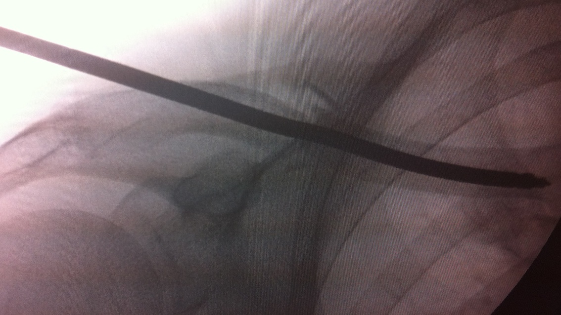
Results
Boehme et al 1991 JBJS Am
- 21 patients established symptomatic non union
- intramedullary Hagie pin
- autologous bone grafting
- 20/21 union
- average time to healing of twenty-two weeks (range, twelve to thirty weeks)
- 17 / 21 the screw had to be removed due to development of a tender bursa
Technique
Open approach to fracture
- 2 - 3 cm
- hand drill medially
- pass cannulated wire laterally and out through skin
- reduce fracture, retrograde pass wire medially
- drill lateral fragment
- insert cannulated 7.3 mm screw
- needs to be between 80 and 110 mm
- check x-ray to ensure good medial fixation
- BG if non union
Post op
- limit ROM above shoulder height for a period
- decreases rotational forces and reduce risk of non union
3. External Fixation
Indications
- open fractures
- severely displaced fractures with damaged skin
Technique
- medial pins are anterior to posterior in an ascending direction to avoid the pleural dome
- lateral pins are superior to inferior in an almost vertical direction
4. Management Malunion
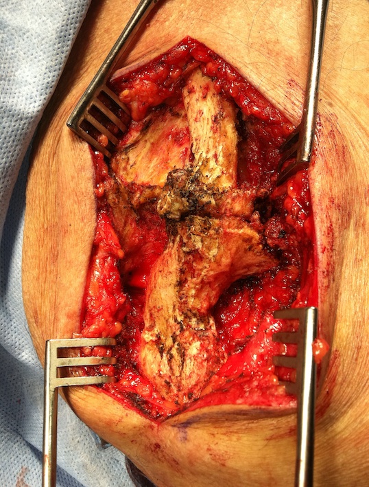
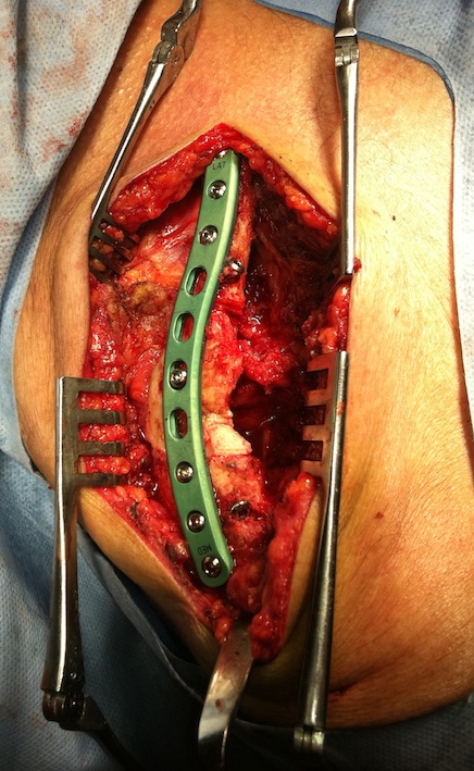
Results
McKee et al JBJS Am 2003
- 15 patients mean age 37 years over 4 year period
- average shortening 2.9 cm
- complaining of pain and fatigueability
- also complaining of symptoms consistent with thoracic outlet syndrome
- many complained of cosmesis
- patients had scapula winging
- osteotomy and DCP (no bone graft required)
- 1 non union
- 8/12 patients with weakness and pain improved
- neurological symptoms eliminated in 7, decreased in 3, unchanged in 1
Complications
Infection / Wound Breakdown
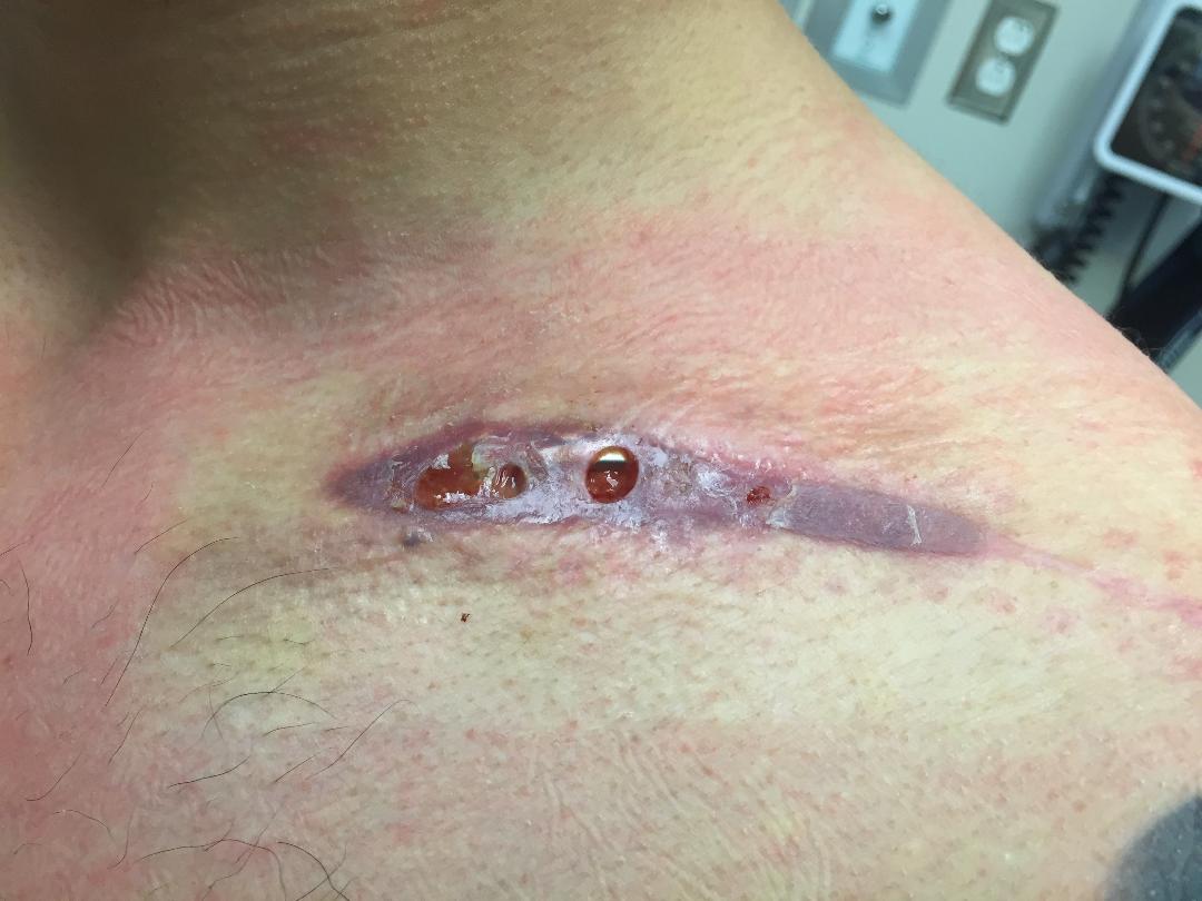
Wound breakdown
- may be a result of vicryl reaction / stitch abcess
- may be better to close fascia over plate, then just use sutures to close wound
Periprosthetic fracture
