Definition
Injury to ulnar collateral ligament of thumb MCPJ
Aetiology
Initial description
- chronic laxity of British gamekeeper's thumb's
- no specific trauma
- secondary to breaking pheasant's neck
Acute trauma
- snow ski
- ball games
Valgus / forced abduction
Anatomy
UCL
- origin medial condyle metacarpal
- passes obliquely volarly
- inserts on volar 1/3 of P1 and volar plate
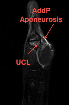
Adductor aponeurosis
- superficial to UCL
- inserts into ulna border thumb extensor mechanism
- via the ulna sesamoid
Examination
Painful, swollen MCPJ
Tenderness along UCL
Abduction Stress Test
- in full extension and 30°
- loss of end point or 30o > other side
- indicates complete rupture
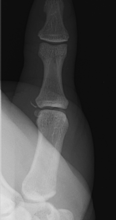
X-ray
3 types Bony avulsion
1. Small fragment pulled away from P1

2. Large intra-articular fracture involving >1/4 articular surface
3. S-H III in paediatric population
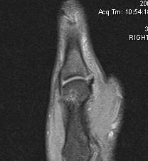
MRI
Look for stenar lesion
- when distal end of UCL
- flipped superficially over adductor aponeurosis
- will not be able to heal
A. Undisplaced


B. Displaced UCL
Management
Non operative
Indications
- partial tear
- undisplaced complete tear
- undisplaced bony fragment
Management
6/52 thumb spica
Operative
Indications for surgery
- complete tear with stener lesion
- large or small displaced bony fragment
- SH III in paediatrics
- chronic injury
Displaced Complete tear / Stener Lesion
Incidence
- 18 - 43%
Types
1. Interposition of the adductor aponeurosis
- between a completely avulsed proximal ulnar collateral ligament injury
- and the proximal phalanx ligament insertion site
2. Interposition between two ends of a mid-substance ligament tear
Diagnosis
Difficult clinically
- may be able to palpate displaced UCL
MRI
Technique
Dorsal incision along ulna border MCPJ
- Divide Adductor pollicis aponeurosis
- leave cuff for lateral repair
- Identify and repair UCL
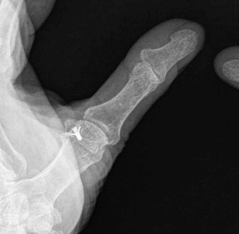
Fixation
- direct suture if able to
- bony anchors
- through drill holes and over lateral button
- cerclage wire
Post op
- 6/52 thumb spica

Bony Avulsion
ORIF Indications
1. > 25% articular surface
2. Small avulsion fracture displaced > 5mm
3. SH III
Chronic Injuries
1. Dynamic tendon transfer
Adductor pollicis
- release adductor pollicis from ulnar sesamoid
- attach to base P1
2. Free tendon graft
Graft options
- palmaris longus
- fourth toe tendon
Technique
- figure of 8 through drill holes
- transverse drill hole base P1
- drill hole head MC
- 6/52 POP
3. Static tendon transfer
EPB
- leave attached distally
- weave through drill holes
