Definition
Pus collection in the epidural space
Epidemiology
Usually haemotogenous seeding
Very rare
- 37 / 1 000 000 patients with LBP
- 1 /10 000 admissions
- most common in old men
Average age 68 years
3/4 males
Rare in paediatrics
Mortality > 12%
Risk Factors
IV drug abuse
Remote infection / UTI
Alcoholics
Invasive spinal procedures / Epidurals
Spinal instrumentation
Immunosuppression
- DM / RA / CRF / Transplant / CA /HIV
Blunt trauma / vertebral fracture
Relhsaus et al Neurosurg Rev 2000
- meta-analysis of 900 cases epidural abscess
- most common risk factors DM / trauma / IVDU / alcoholism
- 5% had had an epidural
- skin infection / abscess most common cause
Pathology
Site
- thoracic spine
- cervical & lumbar spine less common
- spans average of 4 vertebrae
May be anterior or posterior to thecal sac
- dorsal thoracolumbar spine
- ventral 2° vertebral OM / more common in cervical spine
Microbiology
- S aureus 60%
- Streptococcus 10%
- E coli 20% (IVDU, UTI)
- TB
- often unknown
Bacterial Route
1. 1/2 Haematogenous
- remote infection
- UTI / Drug abuse
2. 1/4 Direct Spread
- vertebral osteomyelitis
- abscess usually anterior
3. Following Spinal instrumentation / Surgery/ Epidural injection
4. Adjacent foci
Abscesses
- psoas / pelvic / retropharyngeal / perinephric
1/4 Unknown
Spinal Cord Injury
1. Direct Compression
- mass effect of pus
- ? causes early symptoms
- pus usually tracks freely in epidural space
2. Vascular Occlusion
- decreased arterial flow or epidural vein thrombosis
- responsible for clinical features later in course
- probably more important
Stages
1. Back pain and fever
2. Radicular irritation
3. Weakness / sensory deficit / sphincter incontinence
4. Paralysis
Clinical Features
Classical triad of
1. Back pain & tenderness
2. Fever
3. Elevated ESR
Symptoms
Back pain is hallmark
- 95% / usually very severe / may have nerve root pain
- develops over 72-96 hours
Cord compression < 50%
- weak / numb / urinary Retention
Signs
Fever
- present in 2/3
- may be absent with chronic abscess or antipyretics
Local Signs
- tenderness
- pain on movement
Neurological deficit
- weakness / sensory loss / urinary retention
- may be ambulatory weak / non ambulatory paralysed
- meningitis
Investigations
ESR
- almost always elevated
- usually~ 100
WCC
- usually elevated
Blood Culture
- often identifies organism
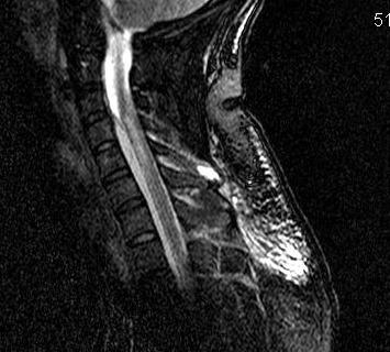
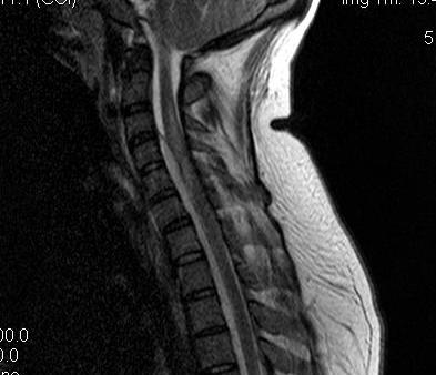
MRI
Investigation of choice
- T1: Low signal intensity mass
- T2: High signal intensity mass
- 85% sensitivity
Gadolinium enhancement T1
- peripherally or homogenous / typical of all abscess on MRI
- increases sensitivity to 95%
Assess levels
- multi level epidural pus
- need multilevel laminectomy and passage of catheter to aid washout
Also assess
- vertebral body osteomyelitis
- cord pathology
- other DDx (HNP, tumour, cord infarct)
Bone Scan
For non specific symptoms
- fever / malaise
- pyrexia of unknown origin (PUO)
- guides further investigation
DDx
Initial diagnosis incorrect in 80% patients
- delayed diagnosis typically
Mechanical LBP
Vertebral OM
Meningitis
Vertebral metastasis
HNP
Transverse Myelitis
Management
Issue
Mainstain of treatment is diagnosis and treatment before neurology develops
- this gives patient best prognosis
Delayed diagnosis most common problem
- 70% patients present with fever and back pain
Poor Prognosis
Delay in diagnosis
Neurology
Cervical / high thoracic
Diabetes
Immunocompromise
Non Operative Management
Indication
Poor surgical candidates
Complete paralysis > 3/7
No neurology
Technique
CT guided biopsy
- obtain cultures / guide antibiotic
- aspiration and drainage of collection
Antibiotics
Treat broad spectrum initially (flucloxacillin + gentamicin)
- 60% S aureus
- 30% Gram negative
- duration of therapy 4 - 8 weeks
Operative Management
Aims
- decompress cord
- debridement / drainage
- MCS of organism
- stabilise spine if needed
Options
1. Posterior laminectomy
- posterior abscess with no anterior body OM
- washout +++
- leave drain in
2. Anterior vertebrectomy and stabilisation
- severe vertebral OM
Prognosis
No significant improvement despite medical advances
Karikari et al Neurosurgery 2009
- 104 patients treated over 10 years
- mortality 17% in non operative / 23% in operative
- 30% with dorsal abscess were quadriplegic / paraplegic
- 7% in the ventral abscess group
- 11% improvement in non operative group
- 25% improvement in operative group