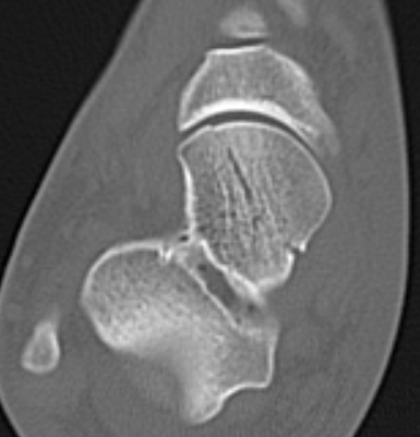
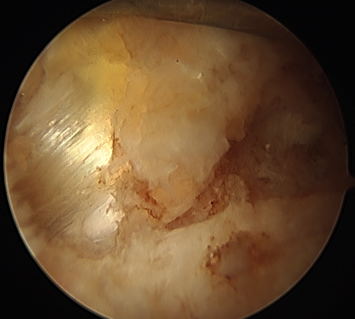
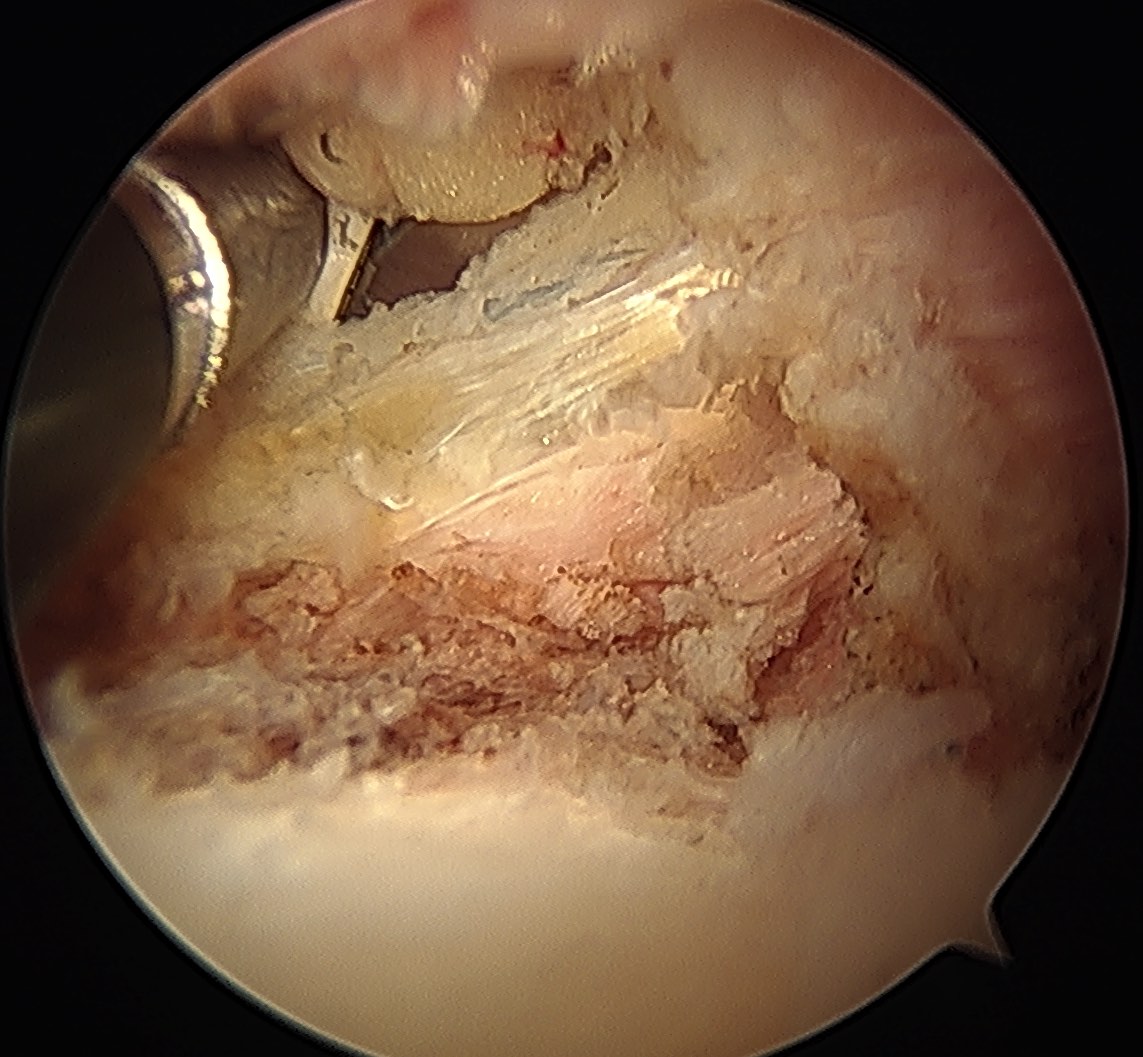
Talar head fractures

Epidemiology
< 10% of all talus fractures
Rare and often missed
Types

< 10% of all talus fractures
Rare and often missed
Superior articular process fracture
- potentially unstable
Inferior articular process fracture
- thought to be more stable


Movement of iliopsoas tendon over femoral head / iliofemoral ridge / iliofemoral ligament
1. Neck of 5th Metacarpal
2. Metacarpal Shaft
3. Metacarpal Head
4. Base of Metacarpal Fracture Dislocations
5. MCPJ dislocations
Non operative Management
Accept 45o angulation
- will have finger extensor lag, but will recover
Facet joint dislocations secondary flexion distraction injury
10%
1. Unifacet subluxation - interspinous process widening
2. Unifacet dislocation - 25% anterolisthesis
3. Bifacet dislocation - 50% anterolisthesis
4. Complete vertebral translation - 100% anterolisthesis
Pathological bone formation in soft tissues
In elbow
- 3% of trauma
- 89% if head injury + trauma
Completely different
1. Myosisitis Ossificans Circumscripta
- post traumatic
- more common
- recognised as a consequence of neurological injury
Knee forms from 3 separate compartments
Plica represents normal embryonic synovial septum that persists into adult life
20% of knees have medial patellar plica at arthroscopy
Symptomatic plicae much less common 1-2%
Mean age 14
Failure of fusion of adjacent ossification centers
Incidence 3%
Bilateral in 60%
4 ossification centers present in acromion
- pre-acromion
- mesoacromion
- metaacromion
- basiacromion
Pre-axial / great toe / 15%
Central / 2-4 MT / 5%
Post-axial / 5th MT / 80%
Type A - articulated
Type B - rudimentary
2 in 1000 births
- 30% positive FHx
- autosomal dominant
Associated MT anomalies common
- block MT / Y-shaped / T-shaped /wide head