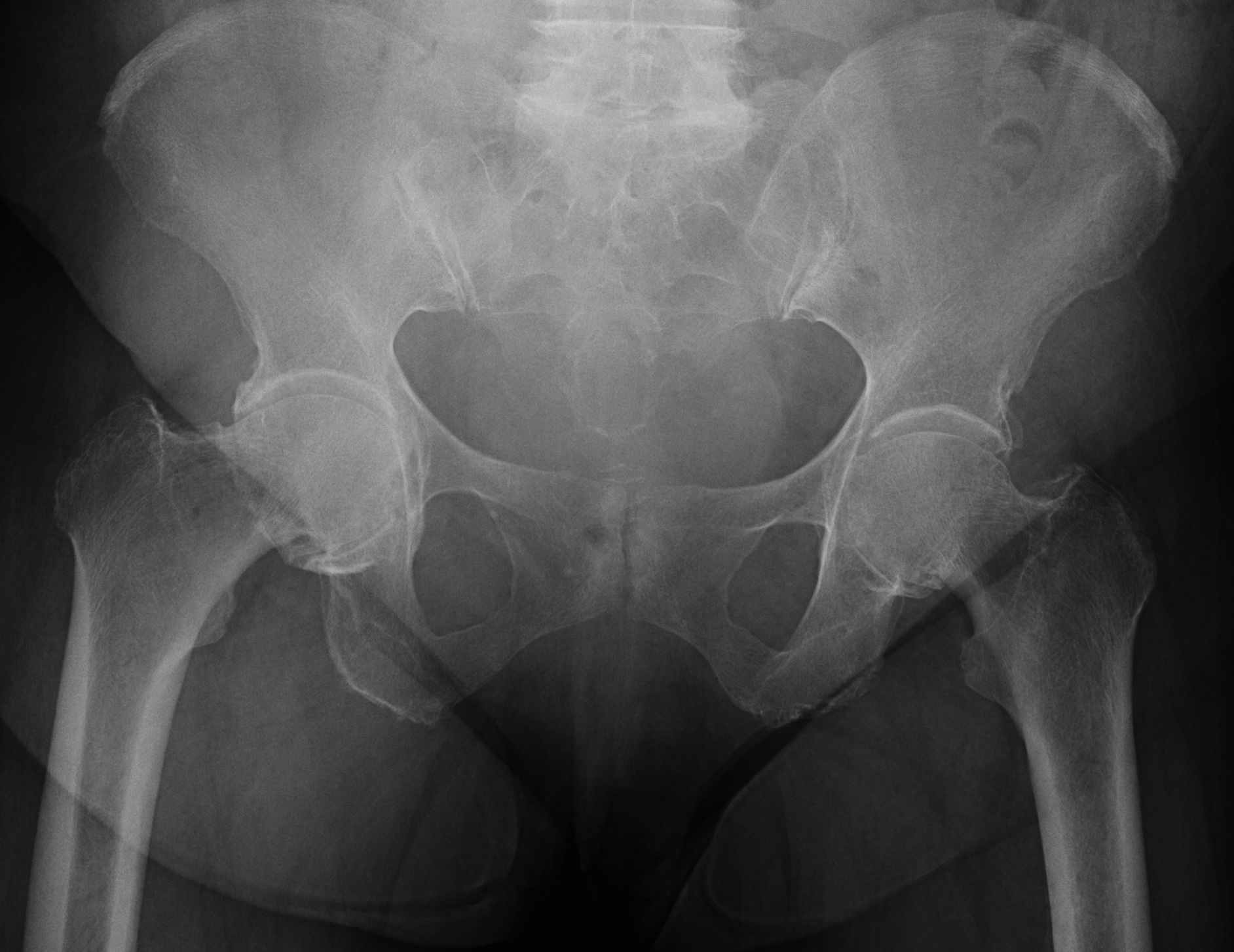
Definition
Deformity of proximal femur with neck-shaft angle <125°
Characterised by
- coxa vara
- decreased femoral anteversion
- limp / trendelenberg
- stress fractures
- early OA
Classification
"ACDDC"
Acquired
2° to underlying disorder
- rickets
- renal osteodystrophy
- hyperparathyroidism
- Perthes disease
- infection
- trauma with early closure physis
- tumour
- SUFE
Congenital Coxa Vara
Dysplasia
- MED
- SED
- Achondroplasia
- Cleidocranial dysostosis
- Fibrous Dysplasia
Developmental / Infantile
- progressive disorder that develops in early childhood
- due to limb bud abnormality
- not congenital really infantile as appears after birth
- includes PFFD & congenitally short femur
Cretinism (hypothyroid)
Developmental Coxa Vara
Epidemiology
- rare
- sex & side incidence equal
- bilateral in 1/3
- increased familial incidence - AD
Aetiology
Unknown / Multifactorial
Theories:
1. Metabolic abnormality - deficiency in proximal femur ossification
2. Excessive intrauterine pressure - causes depression in femoral neck
3. Non-specific mechanical abnormality - occurs during development
4. Vascular insult - arrest in neck development
5. Localized dysplasia - faulty maturation of cartilage & bone in femoral neck
Histology
Abnormality in medial proximal physis & adjacent neck
- cartilage
- 2° metaphyseal bone
Abnormality characterised by
- increased width of physis
- loss of progress of columns
- nests of cartilage in metaphysis
- porotic metaphyseal bone
NHx
Mild
- epiphyseal angle < 45°
- corrects spontaneously
Severe
- epiphyseal angle > 60°
- neck - shaft angle < 110o

Issues
1. Limp / trendelenberg
2. Stress fracture of femoral neck
3. Early degenerative changes
- untreated get severe early OA & often require THR early
Symptoms
Present at walking age with abnormal gait
- painless limp
Signs
Patient is short with hyperlordosis of spine & waddling gait
- limb-length discrepancy
- trendelenburg sign
- mimic DDH
Gait
- short-leg
- trendelenburg sag
- abductor lurch
- if bilateral - waddling gait
Decreased ROM
- especially abduction & IR
Radiology
Inverted Y
- inferior sclerotic metaphyseal triangle
- pathognomonic of developmental
Varus femoral neck
- neck-shaft angle < 125° (normal is 150° in infant)
- difficult to define with severe disease
Hilgenreiner's Epiphyseal angle
- angle between Hilgenreiner's & Physeal line
- normal < 25°
- < 45° should resolve
- 45-60° - watch
- > 60° will progress
Also
- decreased femoral anteversion / retroversion
- coxa breva
Management
Goals
Correction of varus angle
Conversion of femoral neck forces from shear to compression
Correction of LLD
Establish correct abductor tension
Management based on Epiphyseal Angle
<45° - no treatment
45-60° - observe
>60° - valgus osteotomy
Operative Indications
1. Epiphyseal Angle > 60°
2. Epiphyseal Angle 45-60° with limp & progression of varus
Aims
Valgus derotation subtrochanteric osteotomy
- need to overcorrect to 150˚
- epiphyseal angle < 40o
- correct to anteversion 10o
Technique
Lateral approach
- K wire in central head
- mark distal and proximal with drill hole for rotation
- open periosteum and protect with homans
- sub-trochanteric osteotomy with saw
- application of 150o Synthes offset locking plate
- need also IR of about 20° at time of osteotomy
May require
- adductor tenotomy
- femoral shortening
- GT transfer
Plaster spica for 6 weeks post-op
If bilateral do about 6 months apart
Complications
1. Loss of correction
- related to undercorrection
2. Premature physeal closure
- related to increased pressure
- seen in 90% cases
3. Greater trochanter overgrowth
- associated with premature physeal closure
4. Acetabular dysplasia
- associated with premature physeal closure and undercorrection
