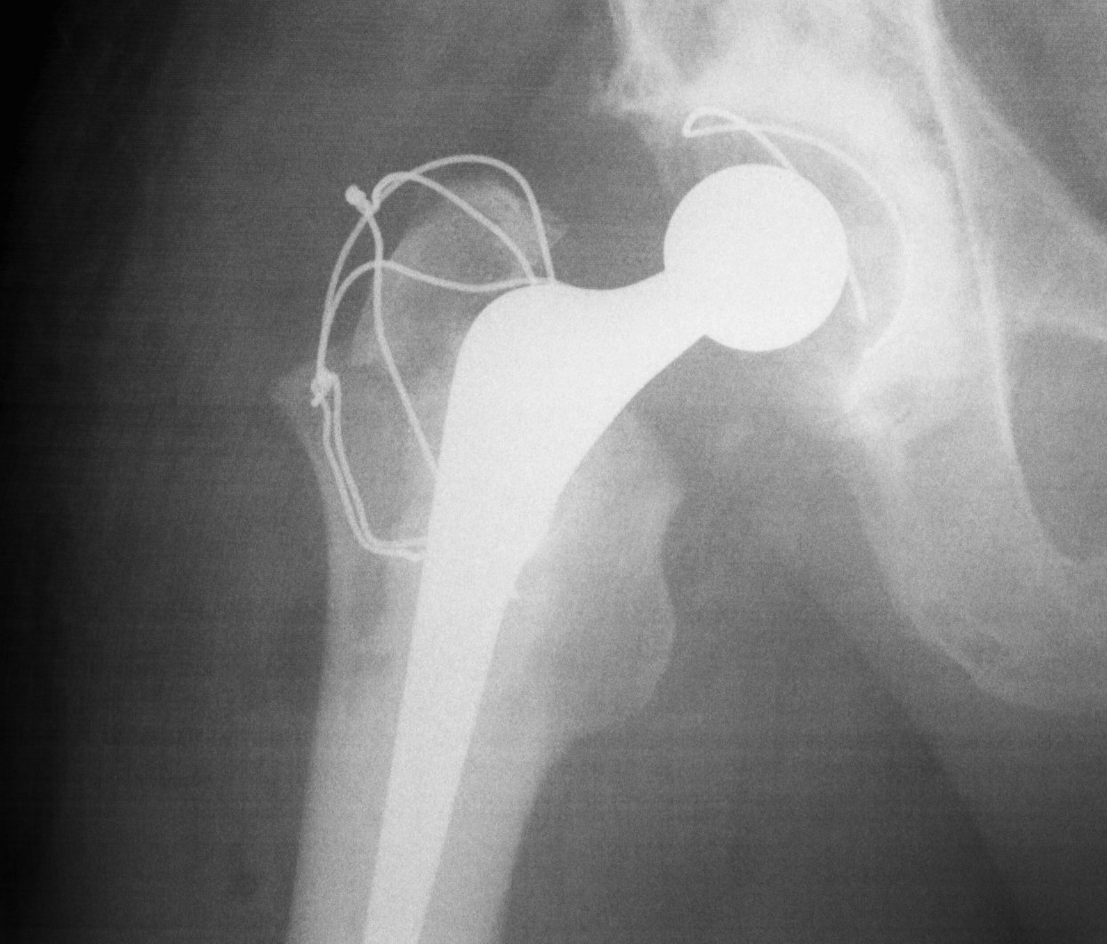
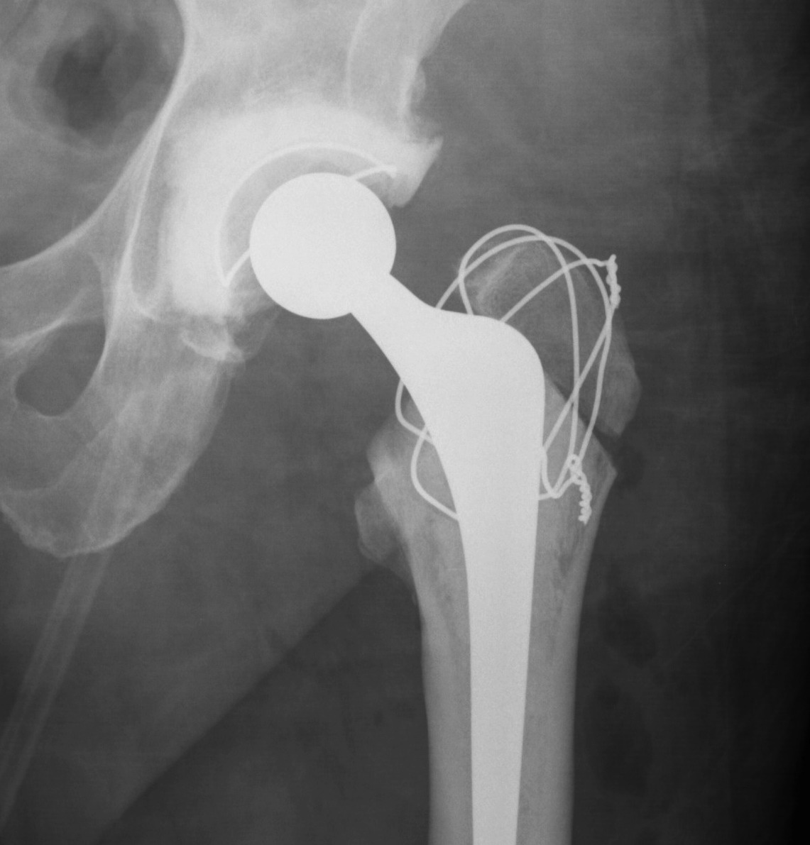
Trochanteric Osteotomy
Types
1. Standard trochanteric osteotomy
2. Sliding trochanteric osteotomy
3. Extended trochanteric osteotomy
Standard Trochanteric osteotomy
1. Standard trochanteric osteotomy
2. Sliding trochanteric osteotomy
3. Extended trochanteric osteotomy
Ranges
- up to 4%
Focal pain
- typically anterolateral thigh
- often tender
- corresponds to tip of stem
1. Instability
Types
- early
- late / failed bony ingrowth
Cause
- micromotion at distal stem
Patient > 70
Gjertsen et al JBJS Am 2010
- 4335 patients > 70 with displaced subcapital fractures
- minimum 1 year follow up
- 1 year mortality same in each group / 25%
- 22% reoperation in ORIF v 3% in hemiarthroplasty
- more pain / higher dissatisfaction / lower quality life in ORIF group
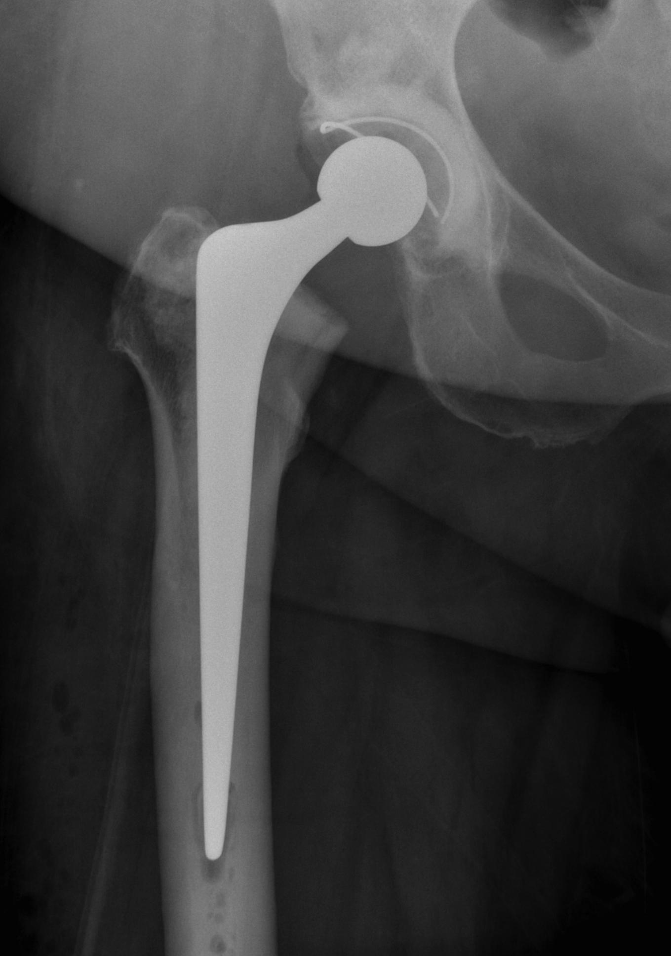
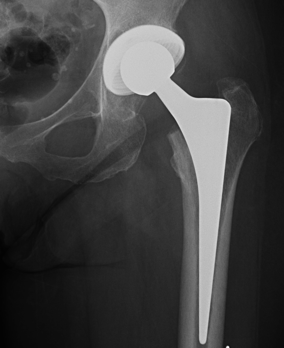
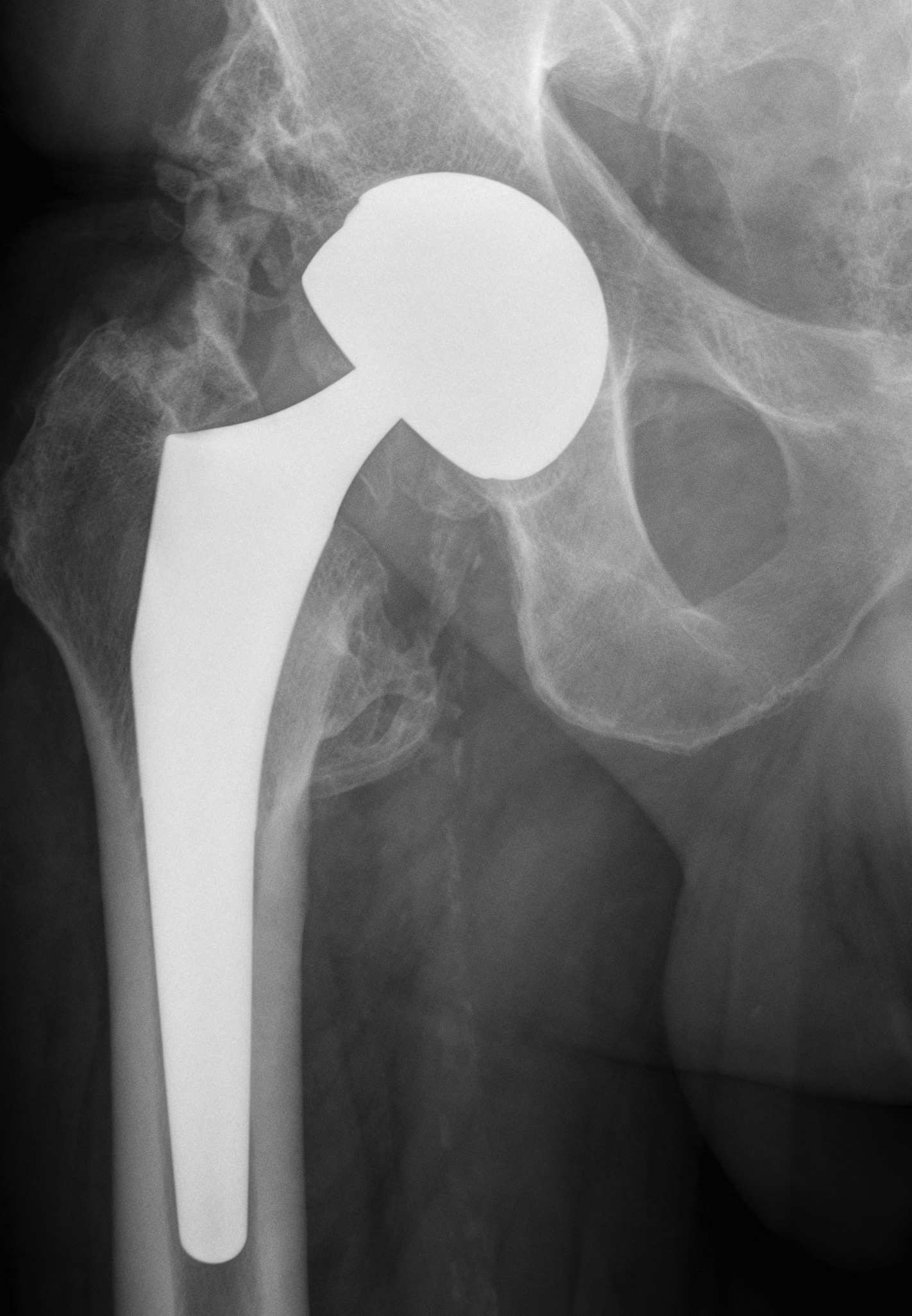
Hemiarthroplasty
- unipolar monoblock
- unipolar modular
Femur
Multiplanar deformity
- worsend by previous surgery
- may require osteotomy
Acetabulum
Dysplasia often present
- not as severe as in DDH
LLD
Can be significant
Abductors
Have been short for long time
- difficult to restore length
Extra-pelvic blood vessels
Femoral Artery
MCFA
LCFA
Profunda Femoris
Obturator artery
Intrapelvic vessels
External iliac artery and vein
Obturator artery
Superior and inferior gluteal
Anatomy
- anterior division of common iliacs / L5-S1
Set up
- on side
- charnley supports posterior on sacrum
- anteriorly on ASIS
- patient slightly tilted backwards
- avoids cup retroversion
Posterior Approach
- identify short ER
Initial press fit
- implant geometry fits the cortical bone in the proximal femur
- good initial mechanical stability
Biological fixation for success
- good press fit
- minimal micromotion
- bony or fibrous tissue ingrowth or ongrowth
Wear
Stability
Normal feel of hip
Increased ROM
Large head
- increase volumetric wear
- less penetrative / linear wear
Small head
- increased linear wear
- decreased volumetric wear
Stage 0
Natural history mixed
- depends on size of lesion and diagnosis
- treat if becomes asymptomatic
- may benefit from bisphosphonates
Stage 1 / Normal X-ray, abnormal MRI
Forage: 80% G/E
Bisphosphonates
Stage 2 / Abnormal X-ray with cysts and sclerosis
A: As for Stage I
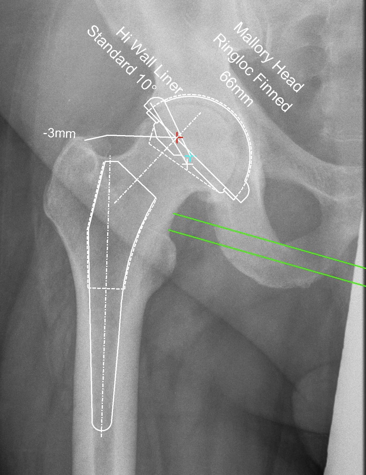
Reproduce the normal anatomical centre of rotation
Restore femoral offset
Maintain equal leg lengths
Usually template off normal hip
1. LLD
2. Offset
3. Femoral component
4. Acetabular component
5. Osteotomy / femoral seating