Epidemiology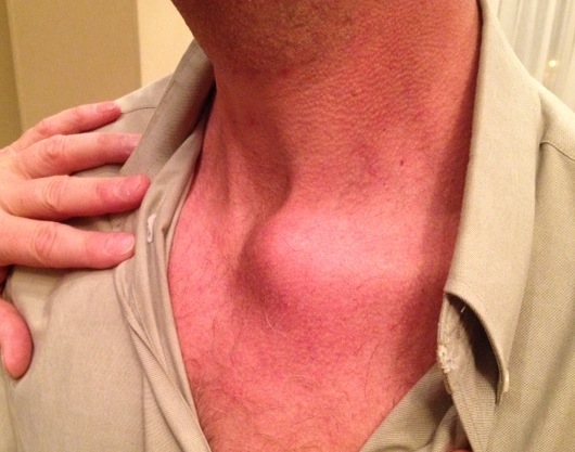
Extremely uncommon
Stability provided by joint capsule /costoclavicular & interclavicular ligaments
Recurrent instability uncommon
Many apparent dislocations in adolescents may be growth plate injuries
-will remodel without treatment
If OA from chronic dislocation may resect SCJ
Types
Anterior & posterior
Posterior
- more serious injury
- least common
Diagnosis
- difficult on physical examination
- radiographs often are non diagnostic
- most consistent diagnostic modality = CT
Anterior
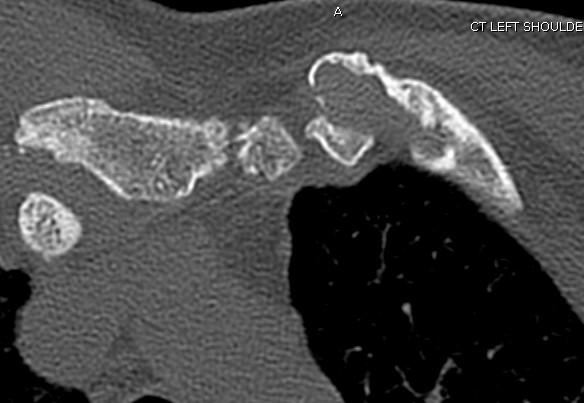
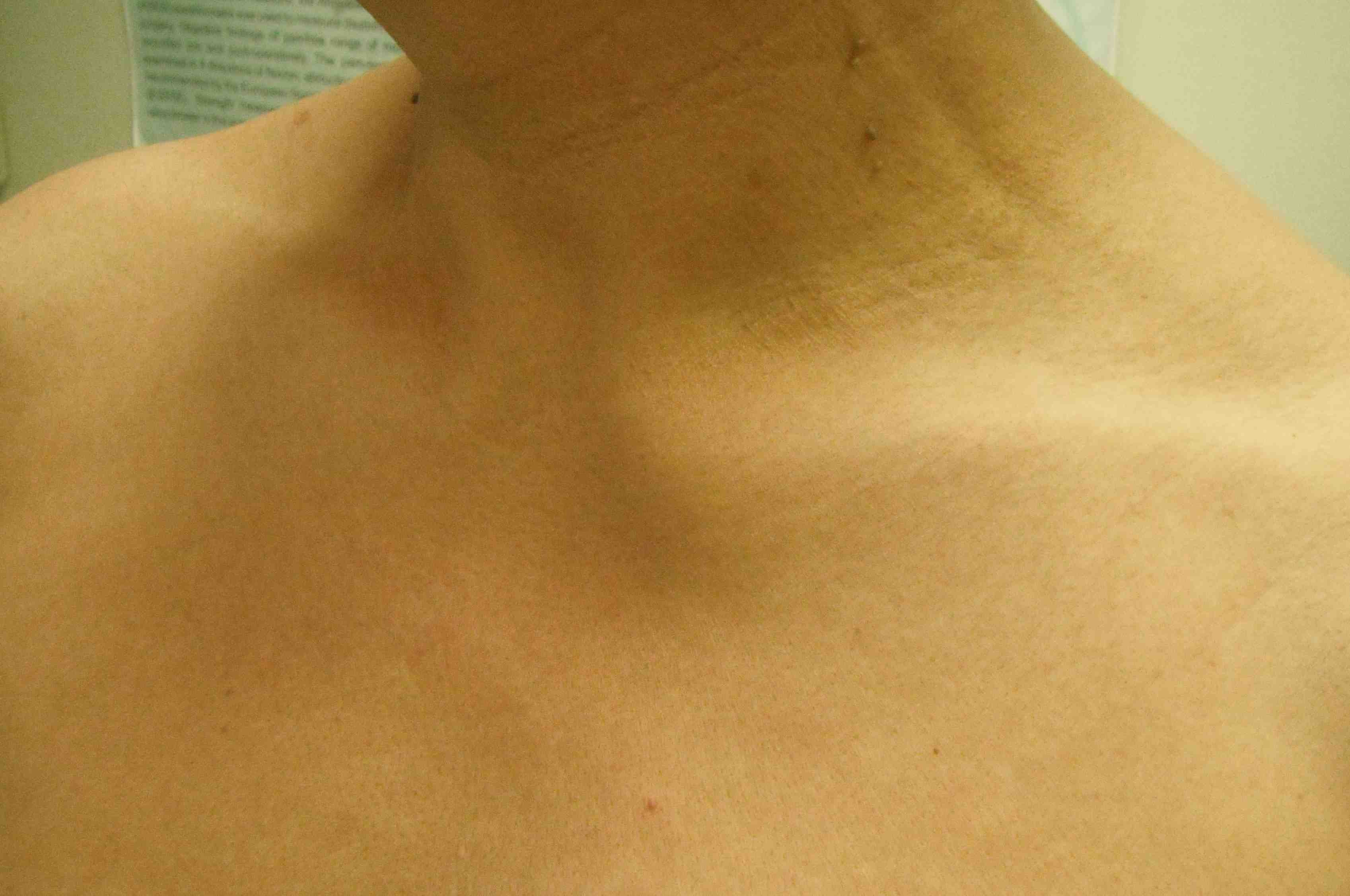
Usually managed non-operatively
- with activity modification and reassurance
MUA
- often will redislocate
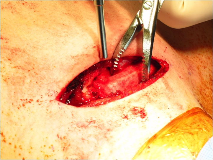
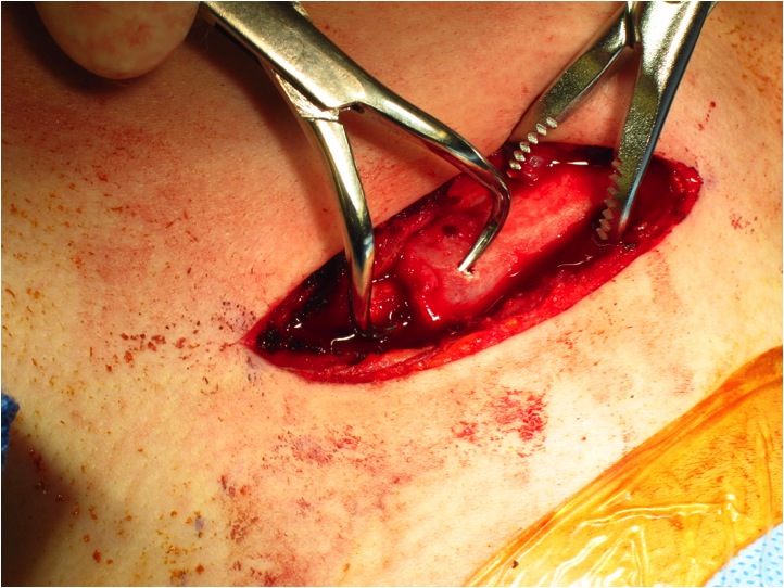
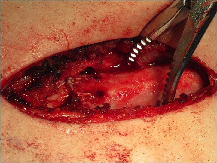
Open reduction
- need to stabilise
- can use strip PL to stabilise
- uncertain if any benefit
Posterior
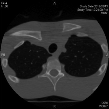
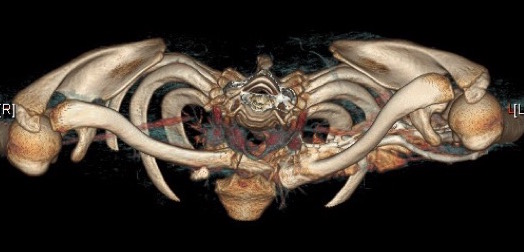
May require treatment because of proximity of major neurovascular structures and airway
1. Closed reduction
- performed under GA in operating room
- chest surgeon available
- potential vascular / airway catastrophe associated with injuries to the mediastinum
- thorough vascular exam pre-operatively
2. Assess stability
Successful closed reduction usually stable
- avoid internal fixation because of likelihood of hardware migration
- possible injury to the mediastinal structures
Closed reduction unsuccessful
- open reduction is indicated
- can stabilize with PL graft / intra-osseous sutures
