Definitions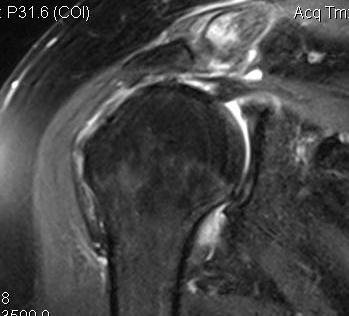
Massive tear
1. > 5cm
- retracted to humerus / glenoid margin
2. At least 2 complete tendons
- lose SS / IS or SS / SC
Classification
Antero-Superior
- SS + SSC
Postero-Superior defects
- SS + IS
- more common
Pathogenesis
Cuff works to compress / depress head in glenoid while deltoid acts as prime mover
- ff still have intact force couple often good function
Plan is to reproduce force couple
- if tear is below equator of head
- get uncoupling of cuff force couple
- lose cuff depressor effect & acts as head elevator
Integrity of coracoacromial arch integral component of repair
- acts as check rein to proximal migration
Presentation
Massive SS / IS wasting + rupture LHB
- weakness
- reduced active ROM
- atrophy
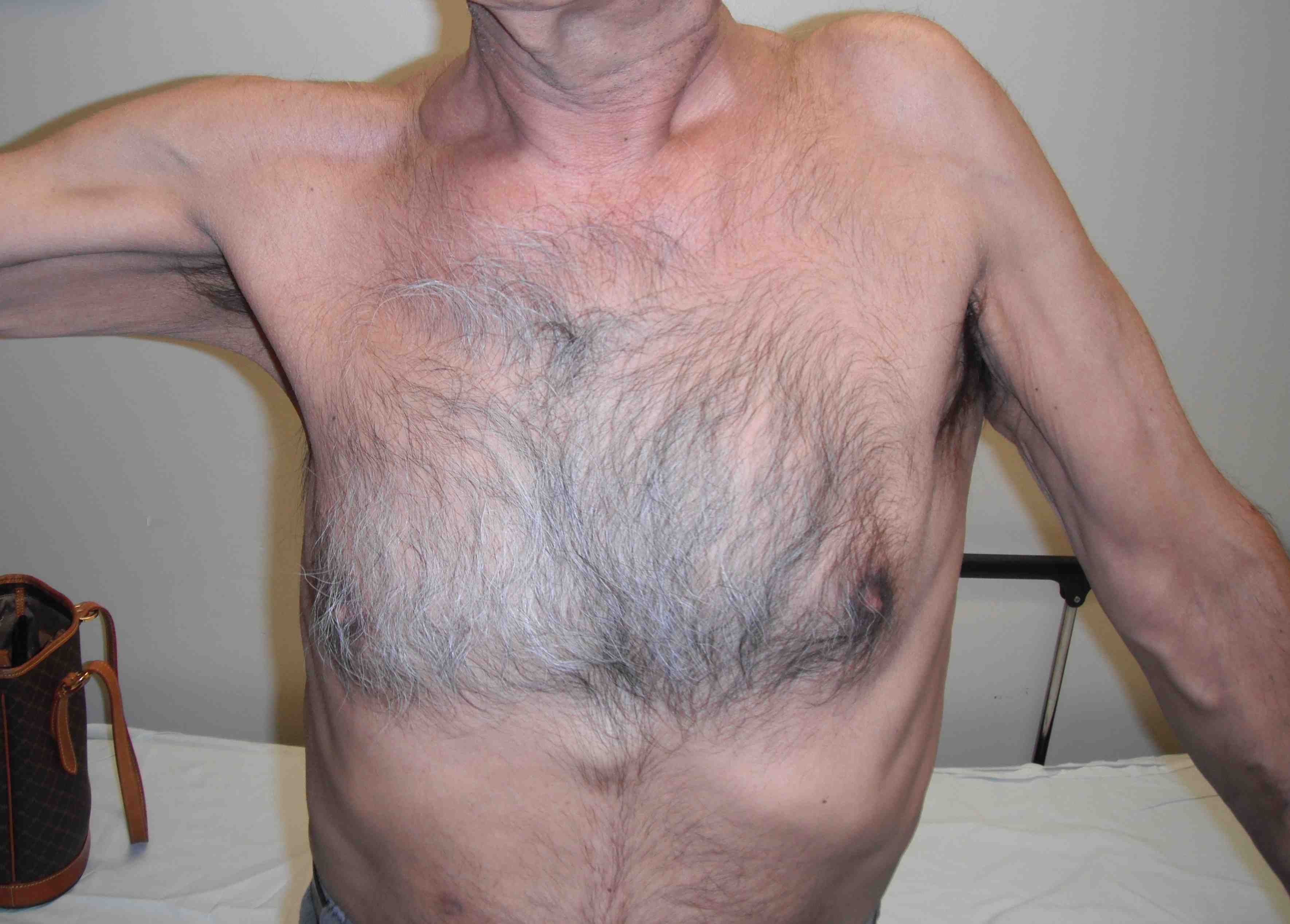
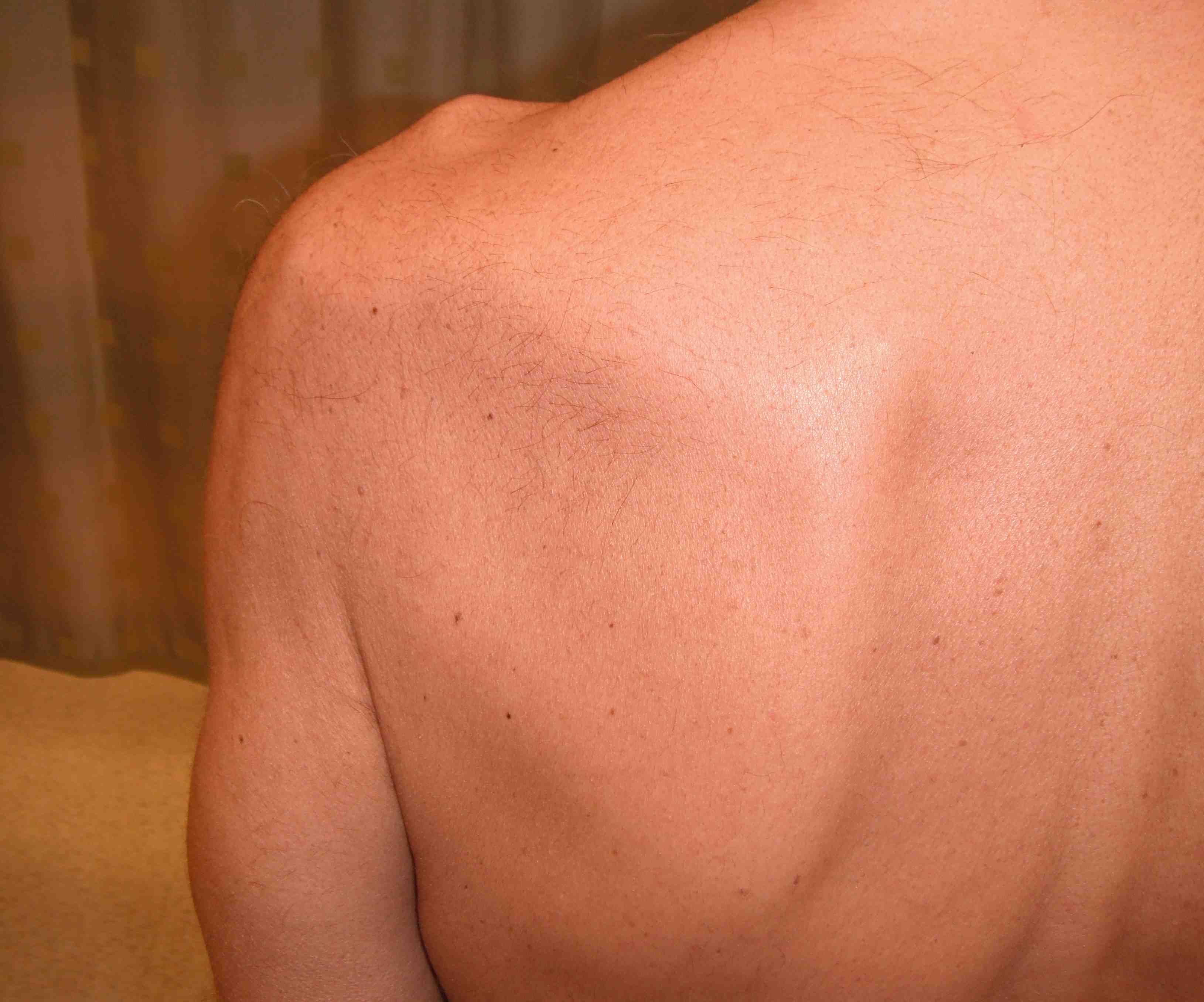
2 classic signs
1. ER lag sign
2. Hornblowers
- 100% sensitive, 93% specific
Both indicate infraspinatous is torn which is usually a sign of a massive PS tear
DDx
Suprascapular nerve palsy
Brachial plexus injury
Cervical stenosis
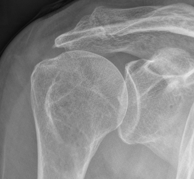
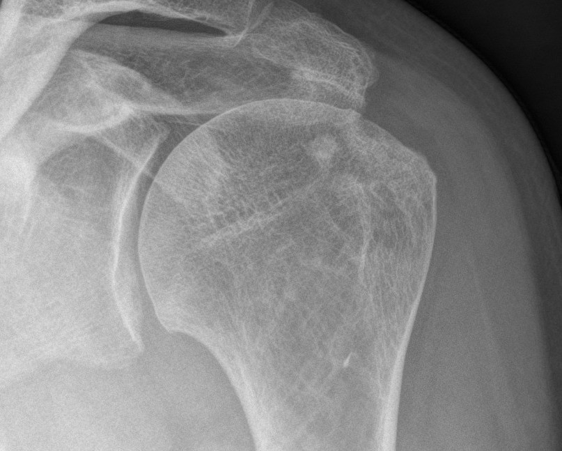
X-ray
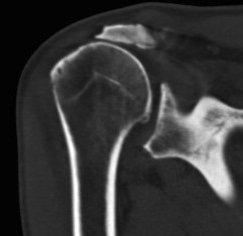
Reduced acromiohumeral space
- < 7 mm RC tear
- < 5 mm massive tear
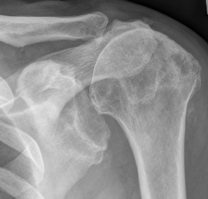
Rotator cuff OA
- acetabularisation
MRI
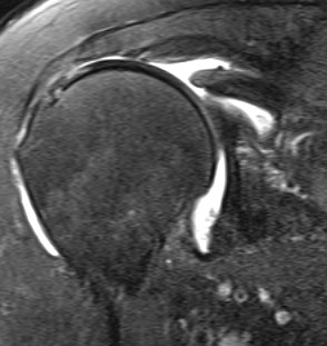
1. Level of retraction
- past coracoid irreparable
2. Quantify fatty infiltration Goutallier
Parasagittal MRI T1
- atrophy and fatty replacement in SS / IS fossa
0 - no fat
1 - minimal fat
2 - more muscle than fat
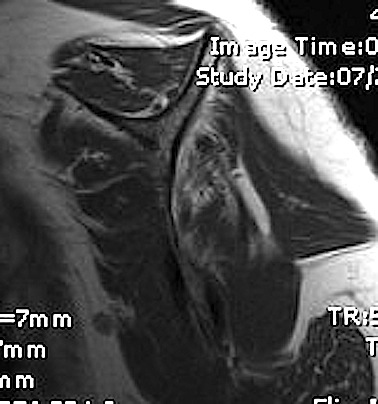
3 - fat equal muscle
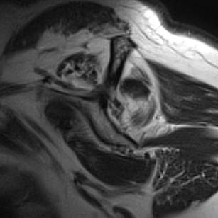
4 - more fat than muscle
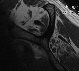
3 & 4 have poor prognosis
- poor functional improvement with repair
- high incidence of retear
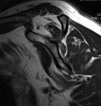
3. Atrophy
Also poor prognosis
Management
Non Operative
Physio / HCLA
- improvement in 50-85%
Operative
Options
A. Primary repair / Debridement
1. Mobilisation and repair
2. Partial repair
3. Decompression and debride
4. Suprascapular nerve release
B. Salvage
1. Local tendon transfer - SSC
2. Distant tendon transfer - P. major / Lat dorsi
3. Allograft
4. Synthetic Graft
5. Arthroplasty
Repair / Debridement
1. Rotator Cuff Mobilisation and repair
Technique of mobilisation
- release coracohumeral ligament
- anterior slide (between SS and SSC)
- posterior slide (between SS and IS)
- release above glenoid 1 cm
- medialise insertion
- transosseous repair
Results
Bigliani et al J Should Elbow Surg 1992
- 61 patients massive cuff tears followed up 7 years
- open repair
- 50% excellent and 30% good
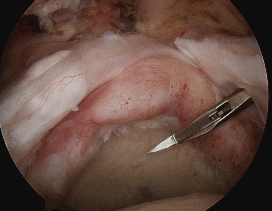
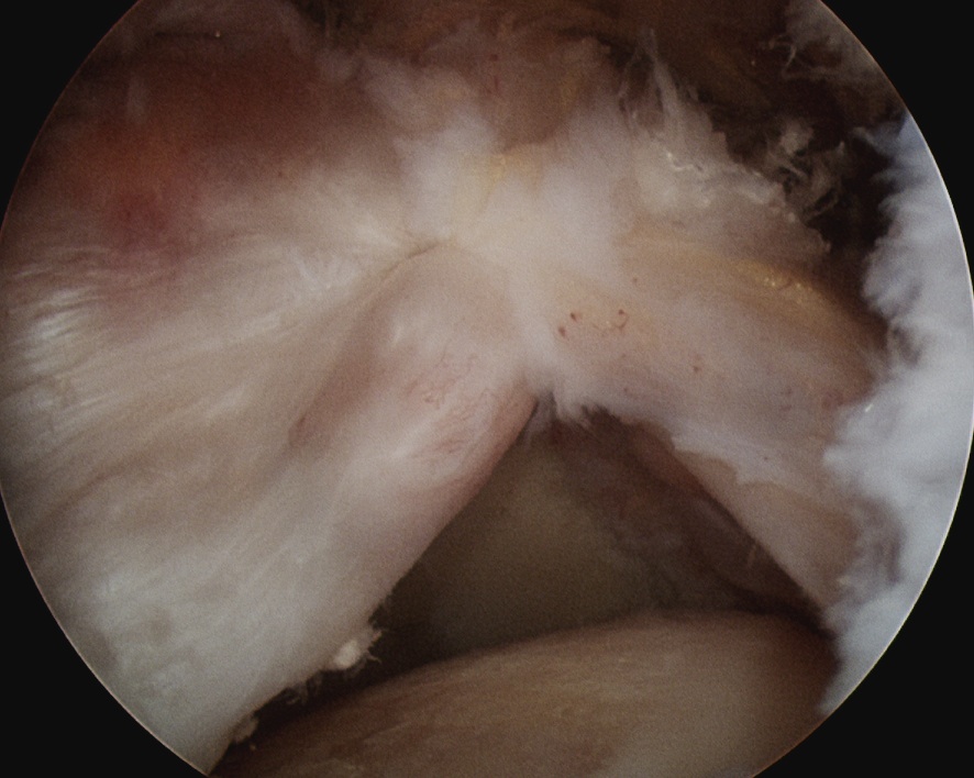
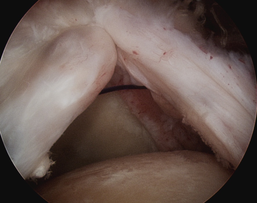
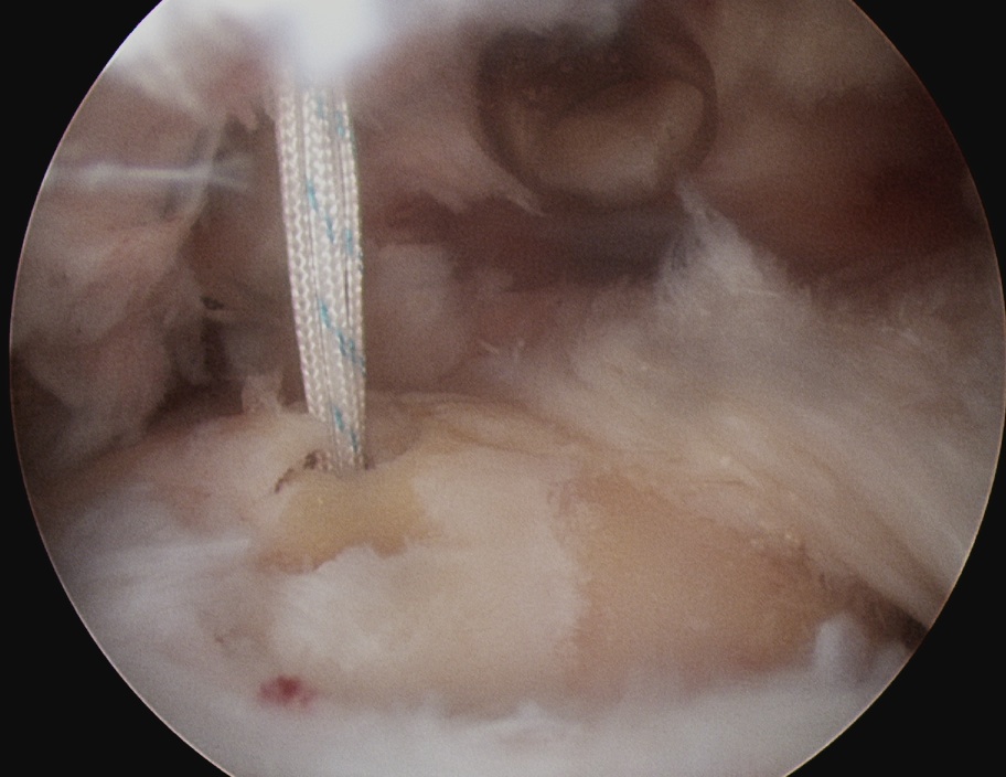
2. Partial repair
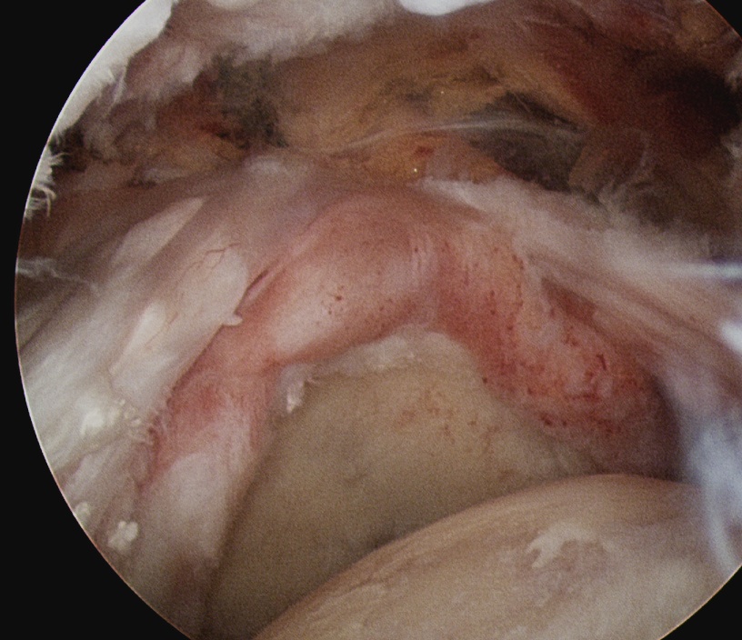
Theory
- restore balanced force couplet
- SSC + partial SS / IS repair
- act in conjuction to depress humeral head
- allow deltoid to work
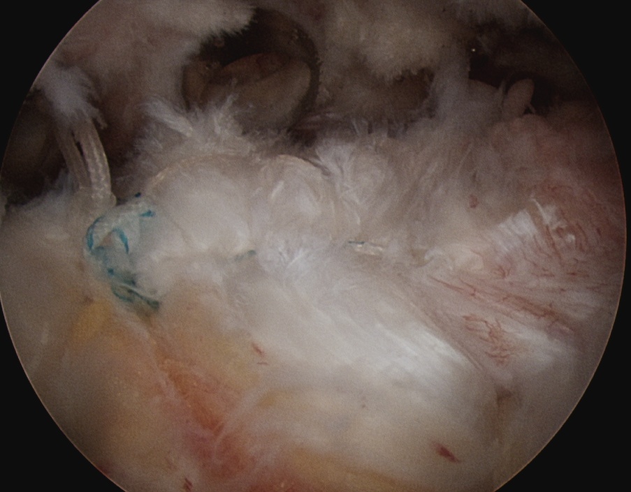
Results
Rhee et al Am J Sports Med 2008
- partial repair with interposition of biceps tendon to bridge gap
- MRI of 14 / 16 cases done arthroscopically
- complete healing in 60%
3. Decompress & debride alone
Concept
- doesn't restore power
- aiming for pain relief in elderly population
Technique
- maintain Coracoacromial arch to prevent humeral head escape
- don't perfrom SAD to preserve CA ligament
- debride cuff edges
- debride GT / tuberoplasty to decrease impingement
- biceps tenotomy / tenodesis
Results
Boileau et al JBJS Am 2007
- demonstrated good results with tenotomy or tenodesis
- 61 patients with irreparable tears
Liem et al Arthroscopy 2008
- 31 patients average age 70
- debridement cuff edges + biceps tenotomy
- no SAD
- reasonable results
Walch et al Arthroscopy 2005
- arthroscopic tenotomy in 307 irreparable RC tears
- 87% satisfied with results
4. Suprascapular nerve release
Theory
- retraction of cuff tethers / impinges SSN
- release of nerve arthroscopically relieves pain
Technique
- arthroscopic release
- see miscellaneous/suprascapular nerve for technique
Salvage
Indications for tendon transfer / Graft
Young patient with poor function
- failed primary repair
- significant weakness
- good deltoid function
- CA arch intact / no superior escape
- good ROM
- either posterosuperior or anterosuperior defect
1. Subscapularis Transfer
Disadvantage
- may lose humeral depressor effect
- lose abduction with deltoid
Technique
- release upper 1/3 tendon from capsule
Results
Karas et al JBJS Am 1996
- 20 patients
- good results in 17
2. P. Major Transfer
Indication
- functional deficit from SSC tear
Technique
- deltopectoral approach
- use sternal head rerouted under clavicular head for better line of pull
Results
Jost et al JBJS Am 2003
- reasonable results in isolated SSC
- less so with combined SS and SSC (doesn't recommend)
3. Lat Dorsii Transfer
Indications
- IS / SS tear
Technique
Lateral Decubitus position
- arm over mayo table
Standard deltoid splitting open approach to subacromial space
- acromioplasty - minimal, preserve CA arch
- ACJ excision if needed
- tag cuff edges medially with sutures to augment repair
- place lateral anchors / sutures
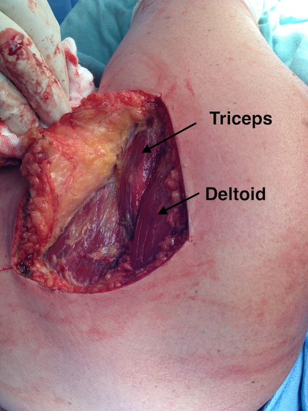
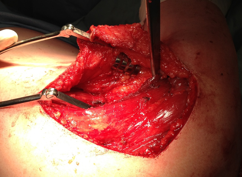
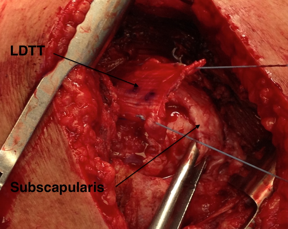
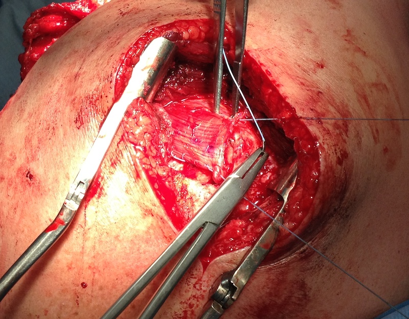
L shaped incision
- inferior margin deltoid, lateral aspect of latissimus dorsi
- arm forward flexed to 90 degrees and IR
- infraspinatous usually very wasted
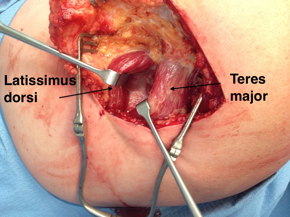
- identify T major
- find L dorsi below T major, develop interval between the two
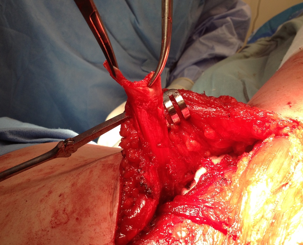
- identify tendon insertion on humerus, often have to release T major tendon from it
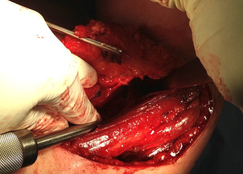
- place homan over humeral head
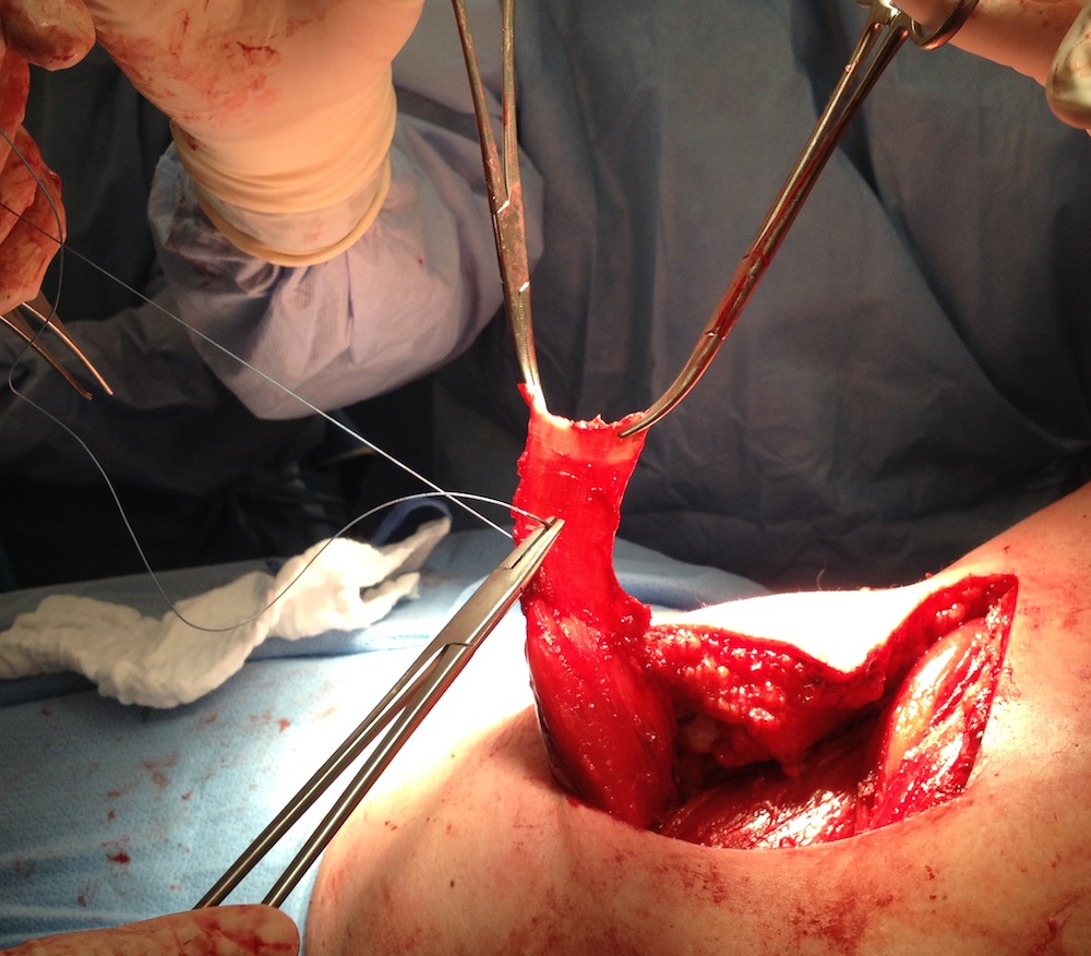
- release tendon from insertion / keep long
- is usually thin / 3 cm wide / 5 cm long
- suture each margin with strong suture, leave limbs long to pass tendon
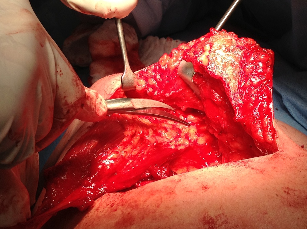
- release muscle belly for length / above and below / must identify and preserve pedicle
- tunnel tendon under deltoid & acromion
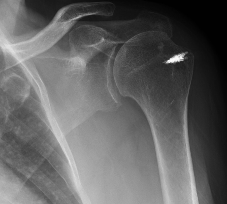
- suture anchors repair to GT + subscapularis + medial cuff remnant
- repair with arm in abduction and ER
- maintain in abduction and external rotation splint for 6/52
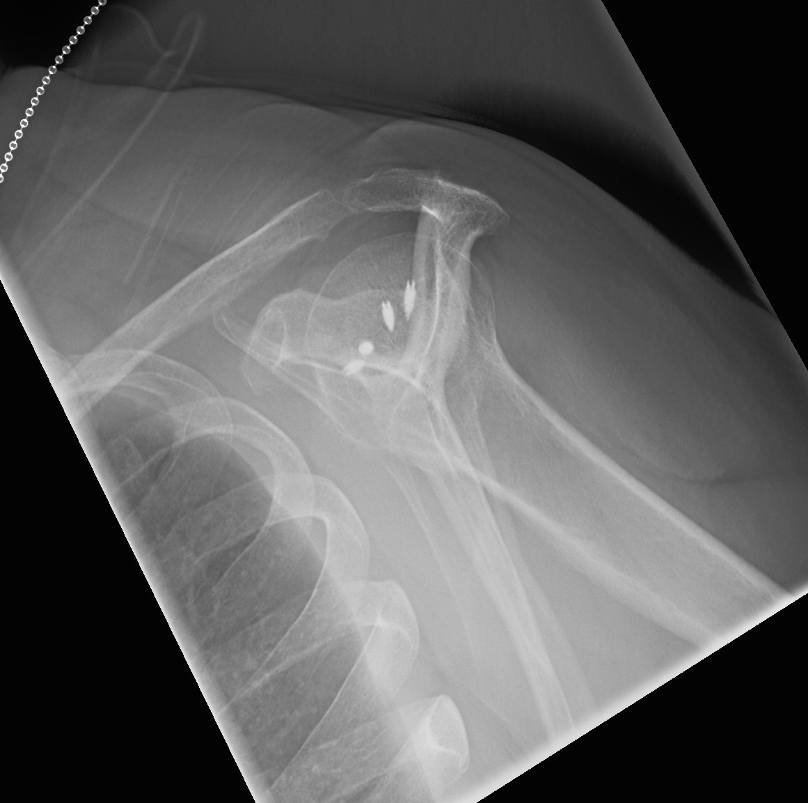
Results
Miniacci JBJS Am 1999
- 14 / 17 good results regarding pain relief and ROM
Tauber et al JBJS Am 2010
- compared patients with tendon transfer to those with tendon + bone block
- significantly improved results in bone block
- 4/22 reruptured on MRI in tendon v 0/20 in bone block group
4. Allograft
Results
Moore et al Am J Sports Med 2006
- 28 patients average age 59
- patella tendon or achilles
- sewn to tendon medially
- bone block laterally or sutured
- 15 repeat MRI - all complete failure of graft
- 1 infection and 1 allograft rejection
- similar functional results to debridement alone
- not recommended by authors
5. Synthetic Allograft
Results
Nada et al JBJS Br 2010
- dacron graft for massive cuff tears in 17 patients
- sutured medially, tied through bony tunnels laterally
- 90% satisfaction
- 15/17 intact on MRI
- 1 rupture, 1 deep infection
6. Arthroplasty
CTA Hemiarthroplasty / Reverse TSR
- salvage in patients > 65 years
